 Movie
Movie Controller

[English] 日本語
 Yorodumi
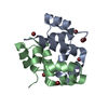
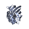
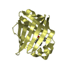
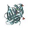
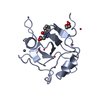
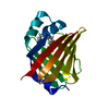
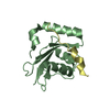
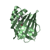
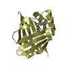
- PDB-3dd7: Structure of DocH66Y in complex with the C-terminal domain of Phd -
+ Open data

- Basic information
| Entry | Database: PDB / ID: 3dd7 | ||||||
|---|---|---|---|---|---|---|---|
| Title | Structure of DocH66Y in complex with the C-terminal domain of Phd | ||||||
 Components Components |
| ||||||
 Keywords Keywords | RIBOSOME INHIBITOR / all alpha | ||||||
| Function / homology |  Function and homology information Function and homology information: / killing by virus of host cell by post-segregational killing / symbiont-mediated suppression of host translation / toxin sequestering activity / DNA-binding transcription repressor activity /  protein-DNA complex / sequence-specific DNA binding / non-specific serine/threonine protein kinase / phosphorylation / protein serine kinase activity ...: / killing by virus of host cell by post-segregational killing / symbiont-mediated suppression of host translation / toxin sequestering activity / DNA-binding transcription repressor activity / protein-DNA complex / sequence-specific DNA binding / non-specific serine/threonine protein kinase / phosphorylation / protein serine kinase activity / protein serine/threonine kinase activity / negative regulation of DNA-templated transcription / protein homodimerization activity / DNA binding / ATP binding protein-DNA complex / sequence-specific DNA binding / non-specific serine/threonine protein kinase / phosphorylation / protein serine kinase activity ...: / killing by virus of host cell by post-segregational killing / symbiont-mediated suppression of host translation / toxin sequestering activity / DNA-binding transcription repressor activity / protein-DNA complex / sequence-specific DNA binding / non-specific serine/threonine protein kinase / phosphorylation / protein serine kinase activity / protein serine/threonine kinase activity / negative regulation of DNA-templated transcription / protein homodimerization activity / DNA binding / ATP bindingSimilarity search - Function | ||||||
| Biological species |  Enterobacteria phage P1 (virus) Enterobacteria phage P1 (virus) | ||||||
| Method | X-RAY DIFFRACTION / SYNCHROTRON / MAD / Resolution: 1.7 Å | ||||||
 Authors Authors | Garcia-Pino, A. / Loris, R. | ||||||
 Citation Citation | Journal: J.Biol.Chem. / Year: 2008 Title: Doc of Prophage P1 Is Inhibited by Its Antitoxin Partner Phd through Fold Complementation Authors: Garcia-Pino, A. / Christensen-Dalsgaard, M. / Wyns, L. / Yarmolinsky, M. / Magnuson, R.D. / Gerdes, K. / Loris, R. | ||||||
| History |
|
- Structure visualization
| Structure viewer | Molecule: MolmilJmol/JSmol |
|---|
- Downloads & links
-Download
| PDBx/mmCIF format | 3dd7.cif.gz | 71.4 KB | Display | PDBx/mmCIF format |
|---|---|---|---|---|
| PDB format | pdb3dd7.ent.gz | 58.2 KB | Display | PDB format |
| PDBx/mmJSON format | 3dd7.json.gz | Tree view | PDBx/mmJSON format | |
| Others |  Other downloads Other downloads |
-Validation report
| Arichive directory | https://data.pdbj.org/pub/pdb/validation_reports/dd/3dd7ftp://data.pdbj.org/pub/pdb/validation_reports/dd/3dd7 | HTTPS FTP |
|---|
-Related structure data
| Related structure data | |
|---|---|
| Similar structure data |


-Links
 PDBj
PDBj

- Assembly
| Deposited unit | 
| ||||||||
|---|---|---|---|---|---|---|---|---|---|
| 1 | 
| ||||||||
| 2 | 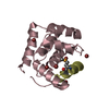
| ||||||||
| Unit cell |
|
-Components
| #1: Protein | Mass: 14782.520 Da / Num. of mol.: 2 / Mutation: H66Y, E126D Source method: isolated from a genetically manipulated source Source: (gene. exp.) Enterobacteria phage P1 (virus) / Gene: doc / Plasmid: pET21b / Production host:  Escherichia coli (E. coli) / Strain (production host): BL21(DE3) / References: UniProt: Q06259 Escherichia coli (E. coli) / Strain (production host): BL21(DE3) / References: UniProt: Q06259#2: Protein/peptide | Mass: 2636.724 Da / Num. of mol.: 2 Fragment: Prevent host death protein C-terminal domain, UNP residues 51-73 Mutation: L70(MSE) / Source method: obtained synthetically / Details: peptide Alta Bioscience / References: UniProt: Q06253 #3: Chemical | ChemComp-BR / Bromide  Mass: 79.904 Da / Num. of mol.: 16 / Source method: obtained synthetically / Formula: Br Mass: 79.904 Da / Num. of mol.: 16 / Source method: obtained synthetically / Formula: Br#4: Water | ChemComp-HOH / | Water Mass: 18.015 Da / Num. of mol.: 228 / Source method: isolated from a natural source / Formula: H2O Mass: 18.015 Da / Num. of mol.: 228 / Source method: isolated from a natural source / Formula: H2O |
|---|
-Experimental details
-Experiment
| Experiment | Method: X-RAY DIFFRACTION / Number of used crystals: 1 |
|---|
- Sample preparation
| Crystal | Density Matthews: 1.91 Å3/Da / Density % sol: 35.71 % |
|---|---|
| Crystal grow | Temperature: 293 K / Method: vapor diffusion, hanging drop / pH: 4.6 Details: 35% PEG4000, 0.2M ammonium acetate, 0.1M acetate, pH4.6, VAPOR DIFFUSION, HANGING DROP, temperature 293.0K |
-Data collection
| Diffraction | Mean temperature: 100 K | |||||||||
|---|---|---|---|---|---|---|---|---|---|---|
| Diffraction source | Source: SYNCHROTRON / Site: EMBL/DESY, HAMBURG  / Beamline: X12 / Wavelength: 0.9189, 0.9116 / Beamline: X12 / Wavelength: 0.9189, 0.9116 | |||||||||
| Detector | Type: MARMOSAIC 225 mm CCD / Detector: CCD / Date: Apr 19, 2007 | |||||||||
| Radiation | Protocol: MAD / Monochromatic (M) / Laue (L): M / Scattering type: x-ray | |||||||||
| Radiation wavelength |
| |||||||||
| Reflection | Resolution: 1.7→11 Å / Num. all: 29371 / Num. obs: 29371 / Observed criterion σ(I): -3 / Redundancy: 7.1 % / Biso Wilson estimate: 19.4 Å2 / Rmerge(I) obs: 0.09 / Net I/σ(I): 9 | |||||||||
| Reflection shell | Resolution: 1.7→1.76 Å / Redundancy: 4.6 % / Rmerge(I) obs: 0.95 / Mean I/σ(I) obs: 2 |
- Processing
| Software |
| |||||||||||||||||||||||||||||||||||||||||||||||||||||||||||||||||||||||||||||||||||||||||||||||||||||||||||||||||||||||||||||||||||||||||||||||||||||||||||||||||||||||||||||||||||||||||||||||||||||||||||||||||||||||||||||||||||||||||||||||||||||||||||||||||||||||||||||||||||||||||||||||||||||||||||||||||||||||||||||||||||||||||||||||||||||||||||||||||||||||||||||||||||||||
|---|---|---|---|---|---|---|---|---|---|---|---|---|---|---|---|---|---|---|---|---|---|---|---|---|---|---|---|---|---|---|---|---|---|---|---|---|---|---|---|---|---|---|---|---|---|---|---|---|---|---|---|---|---|---|---|---|---|---|---|---|---|---|---|---|---|---|---|---|---|---|---|---|---|---|---|---|---|---|---|---|---|---|---|---|---|---|---|---|---|---|---|---|---|---|---|---|---|---|---|---|---|---|---|---|---|---|---|---|---|---|---|---|---|---|---|---|---|---|---|---|---|---|---|---|---|---|---|---|---|---|---|---|---|---|---|---|---|---|---|---|---|---|---|---|---|---|---|---|---|---|---|---|---|---|---|---|---|---|---|---|---|---|---|---|---|---|---|---|---|---|---|---|---|---|---|---|---|---|---|---|---|---|---|---|---|---|---|---|---|---|---|---|---|---|---|---|---|---|---|---|---|---|---|---|---|---|---|---|---|---|---|---|---|---|---|---|---|---|---|---|---|---|---|---|---|---|---|---|---|---|---|---|---|---|---|---|---|---|---|---|---|---|---|---|---|---|---|---|---|---|---|---|---|---|---|---|---|---|---|---|---|---|---|---|---|---|---|---|---|---|---|---|---|---|---|---|---|---|---|---|---|---|---|---|---|---|---|---|---|---|---|---|---|---|---|---|---|---|---|---|---|---|---|---|---|---|---|---|---|---|---|---|---|---|---|---|---|---|---|---|---|---|---|---|---|---|---|---|---|---|---|---|---|---|---|---|---|---|---|---|---|---|---|---|---|---|---|---|---|---|---|---|---|---|---|---|---|---|---|---|---|---|---|---|---|---|---|---|---|---|---|---|---|---|---|---|
| Refinement | Method to determine structure: MAD / Resolution: 1.7→11 Å / Cor.coef. Fo:Fc: 0.959 / Cor.coef. Fo:Fc free: 0.957 / SU B: 3.485 / SU ML: 0.069 / TLS residual ADP flag: LIKELY RESIDUAL / Cross valid method: THROUGHOUT / σ(F): 0 / ESU R: 0.121 / ESU R Free: 0.105 Stereochemistry target values: MAXIMUM LIKELIHOOD WITH PHASES Details: HYDROGENS HAVE BEEN ADDED IN THE RIDING POSITIONS
| |||||||||||||||||||||||||||||||||||||||||||||||||||||||||||||||||||||||||||||||||||||||||||||||||||||||||||||||||||||||||||||||||||||||||||||||||||||||||||||||||||||||||||||||||||||||||||||||||||||||||||||||||||||||||||||||||||||||||||||||||||||||||||||||||||||||||||||||||||||||||||||||||||||||||||||||||||||||||||||||||||||||||||||||||||||||||||||||||||||||||||||||||||||||
| Solvent computation | Ion probe radii: 0.8 Å / Shrinkage radii: 0.8 Å / VDW probe radii: 1.2 Å / Solvent model: MASK | |||||||||||||||||||||||||||||||||||||||||||||||||||||||||||||||||||||||||||||||||||||||||||||||||||||||||||||||||||||||||||||||||||||||||||||||||||||||||||||||||||||||||||||||||||||||||||||||||||||||||||||||||||||||||||||||||||||||||||||||||||||||||||||||||||||||||||||||||||||||||||||||||||||||||||||||||||||||||||||||||||||||||||||||||||||||||||||||||||||||||||||||||||||||
| Displacement parameters | Biso mean: 21.801 Å2
| |||||||||||||||||||||||||||||||||||||||||||||||||||||||||||||||||||||||||||||||||||||||||||||||||||||||||||||||||||||||||||||||||||||||||||||||||||||||||||||||||||||||||||||||||||||||||||||||||||||||||||||||||||||||||||||||||||||||||||||||||||||||||||||||||||||||||||||||||||||||||||||||||||||||||||||||||||||||||||||||||||||||||||||||||||||||||||||||||||||||||||||||||||||||
| Refinement step | Cycle: LAST / Resolution: 1.7→11 Å
| |||||||||||||||||||||||||||||||||||||||||||||||||||||||||||||||||||||||||||||||||||||||||||||||||||||||||||||||||||||||||||||||||||||||||||||||||||||||||||||||||||||||||||||||||||||||||||||||||||||||||||||||||||||||||||||||||||||||||||||||||||||||||||||||||||||||||||||||||||||||||||||||||||||||||||||||||||||||||||||||||||||||||||||||||||||||||||||||||||||||||||||||||||||||
| Refine LS restraints |
| |||||||||||||||||||||||||||||||||||||||||||||||||||||||||||||||||||||||||||||||||||||||||||||||||||||||||||||||||||||||||||||||||||||||||||||||||||||||||||||||||||||||||||||||||||||||||||||||||||||||||||||||||||||||||||||||||||||||||||||||||||||||||||||||||||||||||||||||||||||||||||||||||||||||||||||||||||||||||||||||||||||||||||||||||||||||||||||||||||||||||||||||||||||||
| LS refinement shell | Resolution: 1.7→1.743 Å / Total num. of bins used: 20
| |||||||||||||||||||||||||||||||||||||||||||||||||||||||||||||||||||||||||||||||||||||||||||||||||||||||||||||||||||||||||||||||||||||||||||||||||||||||||||||||||||||||||||||||||||||||||||||||||||||||||||||||||||||||||||||||||||||||||||||||||||||||||||||||||||||||||||||||||||||||||||||||||||||||||||||||||||||||||||||||||||||||||||||||||||||||||||||||||||||||||||||||||||||||
| Refinement TLS params. | Method: refined / Refine-ID: X-RAY DIFFRACTION
| |||||||||||||||||||||||||||||||||||||||||||||||||||||||||||||||||||||||||||||||||||||||||||||||||||||||||||||||||||||||||||||||||||||||||||||||||||||||||||||||||||||||||||||||||||||||||||||||||||||||||||||||||||||||||||||||||||||||||||||||||||||||||||||||||||||||||||||||||||||||||||||||||||||||||||||||||||||||||||||||||||||||||||||||||||||||||||||||||||||||||||||||||||||||
| Refinement TLS group |
|
