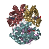
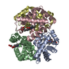
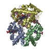
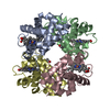
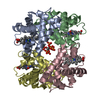
- PDB-3bom: Crystal structure of trout hemoglobin at 1.35 Angstrom resolution -
+
Open data
ID or keywords:
Loading...
-
Basic information
Entry
Database: PDB / ID: 3bom
Title
Crystal structure of trout hemoglobin at 1.35 Angstrom resolution
Components
Hemoglobin subunit alpha-4
Hemoglobin subunit beta-4
Keywords
OXYGEN STORAGE/TRANSPORT / Fish Hemoglobin / Structural Genomics Community Request / Protein Structure Initiative / PSI-2 / Center for Eukaryotic Structural Genomics / CESG / Heme / Iron / Metal-binding / Oxygen transport / Transport / OXYGEN STORAGE-TRANSPORT complex
Mass: 18.015 Da / Num. of mol.: 817 / Source method: isolated from a natural source / Formula: H2O
Has protein modification
Y
-
Experimental details
-
Experiment
Experiment
Method: X-RAY DIFFRACTION / Number of used crystals: 1
-
Sample preparation
Crystal
Density Matthews: 2.2 Å3/Da / Density % sol: 44.7 %
Crystal grow
Temperature: 277 K / Method: vapor diffusion, hanging drop / pH: 5.7 Details: Protein solution (15 mg/mL CO-bound trout IV Hemoglobin, 0.025 M Sodium chloride, 0.010 M Tris-HCl pH 8.0) was mixed one to one with carbon monoxide flushed well solution to yield a final ...Details: Protein solution (15 mg/mL CO-bound trout IV Hemoglobin, 0.025 M Sodium chloride, 0.010 M Tris-HCl pH 8.0) was mixed one to one with carbon monoxide flushed well solution to yield a final concentration of 22.5% PEG 1500 and 0.04-0.06 M MES/Acetate at pH 5.7. The solutions were pH-ed to verify that the crystallization conditions were at pH 5.7 due to the lower buffer molarity. Crystals were cryo-protected with 22.5% PEG 1500, 22.5% Ethylene glycol, 0.04-0.06 M MES/Acetate at pH 5.7 in a single step, VAPOR DIFFUSION, HANGING DROP, temperature 277K
In the structure databanks used in Yorodumi, some data are registered as the other names, "COVID-19 virus" and "2019-nCoV". Here are the details of the virus and the list of structure data.
Jan 31, 2019. EMDB accession codes are about to change! (news from PDBe EMDB page)
EMDB accession codes are about to change! (news from PDBe EMDB page)
The allocation of 4 digits for EMDB accession codes will soon come to an end. Whilst these codes will remain in use, new EMDB accession codes will include an additional digit and will expand incrementally as the available range of codes is exhausted. The current 4-digit format prefixed with “EMD-” (i.e. EMD-XXXX) will advance to a 5-digit format (i.e. EMD-XXXXX), and so on. It is currently estimated that the 4-digit codes will be depleted around Spring 2019, at which point the 5-digit format will come into force.
The EM Navigator/Yorodumi systems omit the EMD- prefix.
Related info.:Q: What is EMD? / ID/Accession-code notation in Yorodumi/EM Navigator
Yorodumi is a browser for structure data from EMDB, PDB, SASBDB, etc.
This page is also the successor to EM Navigator detail page, and also detail information page/front-end page for Omokage search.
The word "yorodu" (or yorozu) is an old Japanese word meaning "ten thousand". "mi" (miru) is to see.
Related info.:EMDB / PDB / SASBDB / Comparison of 3 databanks / Yorodumi Search / Aug 31, 2016. New EM Navigator & Yorodumi / Yorodumi Papers / Jmol/JSmol / Function and homology information / Changes in new EM Navigator and Yorodumi
 Movie
Movie Controller

 Yorodumi
 Open data
 Basic information
 Components
 Keywords
 Function and homology information
 X-RAY DIFFRACTION /
  Authors
 Citation
 Structure visualization
 Downloads & links
 Other downloads
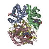


 PDBj
PDBj


 Assembly
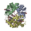

 Mass: 616.487 Da / Num. of mol.: 4 / Source method: obtained synthetically / Formula: C34H32FeN4O4
 Mass: 62.068 Da / Num. of mol.: 4 / Source method: obtained synthetically / Formula: C2H6O2
 Mass: 18.015 Da / Num. of mol.: 817 / Source method: isolated from a natural source / Formula: H2O
 Sample preparation
 / Beamline: 23-ID-D / Wavelength: 0.97935 Å
 Processing