 Movie
Movie Controller


+ Open data

- Basic information
| Entry | Database: PDB / ID: 3anw | ||||||
|---|---|---|---|---|---|---|---|
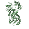
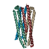
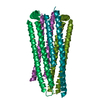
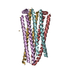
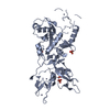
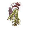
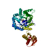
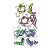
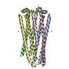
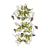
| Title | A protein complex essential initiation of DNA replication | ||||||
 Components Components | (Putative uncharacterized protein) x 2 | ||||||
 Keywords Keywords | REPLICATION / sld5 superfamily / DNA replication | ||||||
| Function / homology |  Function and homology information Function and homology informationArchaeal GINS complex, Gins51 subunit / : / Gins51, C-terminal domain / Methane Monooxygenase Hydroxylase; Chain G, domain 1 - #2050 / Methane Monooxygenase Hydroxylase; Chain G, domain 1 - #1030 / Ribosomal Protein L9; domain 1 - #50 / Ribosomal Protein L9; domain 1 / GINS complex, subunit Psf3 superfamily / GINS subunit, domain A / GINS complex protein helical bundle domain ...Archaeal GINS complex, Gins51 subunit / : / Gins51, C-terminal domain / Methane Monooxygenase Hydroxylase; Chain G, domain 1 - #2050 / Methane Monooxygenase Hydroxylase; Chain G, domain 1 - #1030 / Ribosomal Protein L9; domain 1 - #50 / Ribosomal Protein L9; domain 1 / GINS complex, subunit Psf3 superfamily / GINS subunit, domain A / GINS complex protein helical bundle domain / Methane Monooxygenase Hydroxylase; Chain G, domain 1 / Up-down Bundle / 3-Layer(aba) Sandwich / Mainly Alpha / Alpha Beta Similarity search - Domain/homology | ||||||
| Biological species |   Thermococcus kodakarensis (archaea) Thermococcus kodakarensis (archaea) | ||||||
| Method |  X-RAY DIFFRACTION / SYNCHROTRON / MIR / Resolution: 2.65 Å X-RAY DIFFRACTION / SYNCHROTRON / MIR / Resolution: 2.65 Å | ||||||
 Authors Authors | Oyama, T. / Shirai, T. / Nagasawa, N. / Ishino, Y. / Morikawa, K. | ||||||
 Citation Citation | Journal: Bmc Biol. / Year: 2011 Title: Architectures of archaeal GINS complexes, essential DNA replication initiation factors Authors: Oyama, T. / Ishino, S. / Fujino, S. / Ogino, H. / Shirai, T. / Mayanagi, K. / Saito, M. / Nagasawa, N. / Ishino, Y. / Morikawa, K. | ||||||
| History |
|
- Structure visualization
| Structure viewer | Molecule: MolmilJmol/JSmol |
|---|
- Downloads & links
-Download
| PDBx/mmCIF format | 3anw.cif.gz | 78.6 KB | Display | PDBx/mmCIF format |
|---|---|---|---|---|
| PDB format | pdb3anw.ent.gz | 58.5 KB | Display | PDB format |
| PDBx/mmJSON format | 3anw.json.gz | Tree view | PDBx/mmJSON format | |
| Others |  Other downloads Other downloads |
-Validation report
| Arichive directory | https://data.pdbj.org/pub/pdb/validation_reports/an/3anwftp://data.pdbj.org/pub/pdb/validation_reports/an/3anw | HTTPS FTP |
|---|
-Related structure data
| Similar structure data |
|---|
-Links
 PDBj
PDBj- Assembly
| Deposited unit | 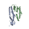
| ||||||||
|---|---|---|---|---|---|---|---|---|---|
| 1 |
| ||||||||
| Unit cell |
|
-Components
| #1: Protein | Mass: 21623.824 Da / Num. of mol.: 1 Source method: isolated from a genetically manipulated source Source: (gene. exp.) Thermococcus kodakarensis (archaea) / Gene: TK0536 / Plasmid: pET28a / Production host:  |
|---|---|
| #2: Protein | Mass: 20029.252 Da / Num. of mol.: 1 Source method: isolated from a genetically manipulated source Source: (gene. exp.) Thermococcus kodakarensis (archaea) / Gene: TK1619 / Plasmid: pET28a / Production host: |
| #3: Water | ChemComp-HOH /  Mass: 18.015 Da / Num. of mol.: 33 / Source method: isolated from a natural source / Formula: H2O Mass: 18.015 Da / Num. of mol.: 33 / Source method: isolated from a natural source / Formula: H2O |
-Experimental details
-Experiment
| Experiment | Method: X-RAY DIFFRACTION / Number of used crystals: 1 |
|---|
- Sample preparation
| Crystal | Density Matthews: 3.17 Å3/Da / Density % sol: 61.22 % |
|---|---|
| Crystal grow | Temperature: 293 K / Method: vapor diffusion, hanging drop / pH: 7.5 Details: 0.1M HEPES, 0.2M magnesium chloride, 24% PEG 400, pH 7.5, VAPOR DIFFUSION, HANGING DROP, temperature 293K |
-Data collection
| Diffraction | Mean temperature: 100 K |
|---|---|
| Diffraction source | Source: SYNCHROTRON / Site: SPring-8  / Beamline: BL38B1 / Wavelength: 1 Å / Beamline: BL38B1 / Wavelength: 1 Å |
| Detector | Type: ADSC QUANTUM 210 / Detector: CCD / Date: Jul 5, 2010 |
| Radiation | Monochromator: FIXED EXIT SI 111 DOUBLE CRYSTAL MONOCHROMATOR Protocol: SINGLE WAVELENGTH / Monochromatic (M) / Laue (L): M / Scattering type: x-ray |
| Radiation wavelength | Wavelength: 1 Å / Relative weight: 1 |
| Reflection | Resolution: 2.65→37.35 Å / Num. all: 16278 / Num. obs: 16278 / % possible obs: 99.4 % / Observed criterion σ(F): 0 / Observed criterion σ(I): 0 / Redundancy: 14 % / Biso Wilson estimate: 55.9 Å2 / Rmerge(I) obs: 0.043 / Net I/σ(I): 17.6 |
| Reflection shell | Resolution: 2.65→2.74 Å / Redundancy: 14 % / Rmerge(I) obs: 0.388 / Mean I/σ(I) obs: 9.9 / Num. unique all: 1598 / % possible all: 100 |
- Processing
| Software |
| ||||||||||||||||||||||||||||||||||||
|---|---|---|---|---|---|---|---|---|---|---|---|---|---|---|---|---|---|---|---|---|---|---|---|---|---|---|---|---|---|---|---|---|---|---|---|---|---|
| Refinement | Method to determine structure: MIR / Resolution: 2.65→37.35 Å / Rfactor Rfree error: 0.011 / Data cutoff high absF: 116289.88 / Data cutoff low absF: 0 / Isotropic thermal model: RESTRAINED / Cross valid method: THROUGHOUT / σ(F): 0 / Stereochemistry target values: Engh & Huber / Details: BULK SOLVENT MODEL USED
| ||||||||||||||||||||||||||||||||||||
| Solvent computation | Solvent model: FLAT MODEL / Bsol: 43.9503 Å2 / ksol: 0.35 e/Å3 | ||||||||||||||||||||||||||||||||||||
| Displacement parameters | Biso mean: 64.8 Å2
| ||||||||||||||||||||||||||||||||||||
| Refine analyze |
| ||||||||||||||||||||||||||||||||||||
| Refinement step | Cycle: LAST / Resolution: 2.65→37.35 Å
| ||||||||||||||||||||||||||||||||||||
| Refine LS restraints |
| ||||||||||||||||||||||||||||||||||||
| Refine LS restraints NCS | NCS model details: NONE | ||||||||||||||||||||||||||||||||||||
| LS refinement shell | Resolution: 2.65→2.82 Å / Rfactor Rfree error: 0.045 / Total num. of bins used: 6
| ||||||||||||||||||||||||||||||||||||
| Xplor file |
|
