 Movie
Movie Controller


+ Open data

- Basic information
| Entry | Database: PDB / ID: 3a7m | ||||||
|---|---|---|---|---|---|---|---|
| Title | Structure of FliT, the flagellar type III chaperone for FliD | ||||||
 Components Components | Flagellar protein fliT | ||||||
 Keywords Keywords | GENE REGULATION / CHAPERONE / Up-down helix bundle / Bacterial flagellum biogenesis / Cytoplasm / Repressor / Transcription / Transcription regulation | ||||||
| Function / homology |  Function and homology information Function and homology informationnegative regulation of bacterial-type flagellum assembly / bacterial-type flagellum organization / protein folding / identical protein binding / cytosol Similarity search - Function | ||||||
| Biological species |  Salmonella typhimurium (bacteria) Salmonella typhimurium (bacteria) | ||||||
| Method |  X-RAY DIFFRACTION / SYNCHROTRON / SAD / Resolution: 3.2 Å X-RAY DIFFRACTION / SYNCHROTRON / SAD / Resolution: 3.2 Å | ||||||
 Authors Authors | Imada, K. / Minamino, T. / Kinoshita, M. / Namba, K. | ||||||
 Citation Citation | Journal: Proc.Natl.Acad.Sci.USA / Year: 2010 Title: Structural insight into the regulatory mechanisms of interactions of the flagellar type III chaperone FliT with its binding partners. Authors: Imada, K. / Minamino, T. / Kinoshita, M. / Furukawa, Y. / Namba, K. | ||||||
| History |
|
- Structure visualization
| Structure viewer | Molecule: MolmilJmol/JSmol |
|---|
- Downloads & links
-Download
| PDBx/mmCIF format | 3a7m.cif.gz | 56.2 KB | Display | PDBx/mmCIF format |
|---|---|---|---|---|
| PDB format | pdb3a7m.ent.gz | 42.7 KB | Display | PDB format |
| PDBx/mmJSON format | 3a7m.json.gz | Tree view | PDBx/mmJSON format | |
| Others |  Other downloads Other downloads |
-Validation report
| Arichive directory | https://data.pdbj.org/pub/pdb/validation_reports/a7/3a7mftp://data.pdbj.org/pub/pdb/validation_reports/a7/3a7m | HTTPS FTP |
|---|
-Related structure data
| Similar structure data |
|---|


-Links
 PDBj
PDBj
- Assembly
| Deposited unit | 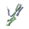
| ||||||||
|---|---|---|---|---|---|---|---|---|---|
| 1 |
| ||||||||
| Unit cell |
| ||||||||
| Details | AUTHOR DEFINED BIOLOGICAL UNIT: UNKNOWN |
-Components
| #1: Protein | Mass: 13903.202 Da / Num. of mol.: 2 Source method: isolated from a genetically manipulated source Source: (gene. exp.) Salmonella typhimurium (bacteria) / Strain: SJW1103 / Gene: fliT, STM1962 / Plasmid: PET3c / Production host: Has protein modification | Y | |
|---|
-Experimental details
-Experiment
| Experiment | Method: X-RAY DIFFRACTION / Number of used crystals: 1 |
|---|
- Sample preparation
| Crystal | Density Matthews: 4.49 Å3/Da / Density % sol: 72.58 % |
|---|---|
| Crystal grow | Temperature: 277 K / Method: vapor diffusion, sitting drop / pH: 9.5 Details: 1M Potassium sodium tartrate, 0.1M CHES, 0.2M Li2SO4, pH9.5, VAPOR DIFFUSION, SITTING DROP, temperature 277K |
-Data collection
| Diffraction | Mean temperature: 35 K / Ambient temp details: He gas cooling cryo system |
|---|---|
| Diffraction source | Source: SYNCHROTRON / Site: SPring-8  / Beamline: BL41XU / Wavelength: 0.97915 Å / Beamline: BL41XU / Wavelength: 0.97915 Å |
| Detector | Type: ADSC QUANTUM 315 / Detector: CCD / Date: Jan 1, 2008 |
| Radiation | Monochromator: Double-crystal monochromator / Protocol: SINGLE WAVELENGTH / Monochromatic (M) / Laue (L): M / Scattering type: x-ray |
| Radiation wavelength | Wavelength: 0.97915 Å / Relative weight: 1 |
| Reflection | Resolution: 3.2→42.2 Å / Num. all: 8390 / Num. obs: 8390 / % possible obs: 99.3 % / Observed criterion σ(F): 0 / Observed criterion σ(I): 0 / Redundancy: 3.9 % / Biso Wilson estimate: 70.1 Å2 / Rsym value: 0.101 / Net I/σ(I): 11.1 |
| Reflection shell | Resolution: 3.2→3.37 Å / Redundancy: 4 % / Mean I/σ(I) obs: 2.6 / Num. unique all: 1219 / Rsym value: 0.402 / % possible all: 99.9 |
- Processing
| Software |
| ||||||||||||||||||||||||||||||||||||||||||||||||||||||||||||||||||||||||||||||||
|---|---|---|---|---|---|---|---|---|---|---|---|---|---|---|---|---|---|---|---|---|---|---|---|---|---|---|---|---|---|---|---|---|---|---|---|---|---|---|---|---|---|---|---|---|---|---|---|---|---|---|---|---|---|---|---|---|---|---|---|---|---|---|---|---|---|---|---|---|---|---|---|---|---|---|---|---|---|---|---|---|---|
| Refinement | Method to determine structure: SAD / Resolution: 3.2→42.19 Å / Rfactor Rfree error: 0.01 / Data cutoff high absF: 1423711.61 / Data cutoff low absF: 0 / Isotropic thermal model: RESTRAINED / Cross valid method: THROUGHOUT / σ(F): 0 / Stereochemistry target values: Engh & Huber
| ||||||||||||||||||||||||||||||||||||||||||||||||||||||||||||||||||||||||||||||||
| Solvent computation | Solvent model: FLAT MODEL / Bsol: 61.7012 Å2 / ksol: 0.403934 e/Å3 | ||||||||||||||||||||||||||||||||||||||||||||||||||||||||||||||||||||||||||||||||
| Displacement parameters | Biso mean: 52.5 Å2
| ||||||||||||||||||||||||||||||||||||||||||||||||||||||||||||||||||||||||||||||||
| Refine analyze |
| ||||||||||||||||||||||||||||||||||||||||||||||||||||||||||||||||||||||||||||||||
| Refinement step | Cycle: LAST / Resolution: 3.2→42.19 Å
| ||||||||||||||||||||||||||||||||||||||||||||||||||||||||||||||||||||||||||||||||
| Refine LS restraints |
| ||||||||||||||||||||||||||||||||||||||||||||||||||||||||||||||||||||||||||||||||
| LS refinement shell | Resolution: 3.2→3.4 Å / Rfactor Rfree error: 0.031 / Total num. of bins used: 6
| ||||||||||||||||||||||||||||||||||||||||||||||||||||||||||||||||||||||||||||||||
| Xplor file |
|
