 Movie
Movie Controller

[English] 日本語
 Yorodumi
- PDB-2zvy: Structure of the periplasmic domain of MotB from Salmonella (crys... -
+ Open data

- Basic information
| Entry | Database: PDB / ID: 2zvy | ||||||
|---|---|---|---|---|---|---|---|
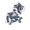
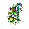
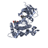
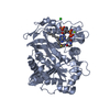
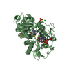
| Title | Structure of the periplasmic domain of MotB from Salmonella (crystal form II) | ||||||
 Components Components | Chemotaxis protein motB | ||||||
 Keywords Keywords | MEMBRANE PROTEIN / 2-layer sandwich / Bacterial flagellum / Cell inner membrane / Cell membrane / Chemotaxis / Flagellar rotation / Membrane / Transmembrane | ||||||
| Function / homology |  Function and homology information Function and homology informationarchaeal or bacterial-type flagellum-dependent cell motility / chemotaxis / plasma membrane Similarity search - Function | ||||||
| Biological species |  Salmonella typhimurium (bacteria) Salmonella typhimurium (bacteria) | ||||||
| Method |  X-RAY DIFFRACTION / SYNCHROTRON / MOLECULAR REPLACEMENT / Resolution: 1.75 Å X-RAY DIFFRACTION / SYNCHROTRON / MOLECULAR REPLACEMENT / Resolution: 1.75 Å | ||||||
 Authors Authors | Kojima, S. / Homma, M. / Namba, K. / Imada, K. | ||||||
 Citation Citation | Journal: Mol.Microbiol. / Year: 2009 Title: Stator assembly and activation mechanism of the flagellar motor by the periplasmic region of MotB Authors: Kojima, S. / Imada, K. / Sakuma, M. / Sudo, Y. / Kojima, C. / Minamino, T. / Homma, M. / Namba, K. | ||||||
| History |
|
- Structure visualization
| Structure viewer | Molecule: MolmilJmol/JSmol |
|---|
- Downloads & links
-Download
| PDBx/mmCIF format | 2zvy.cif.gz | 94.2 KB | Display | PDBx/mmCIF format |
|---|---|---|---|---|
| PDB format | pdb2zvy.ent.gz | 71.2 KB | Display | PDB format |
| PDBx/mmJSON format | 2zvy.json.gz | Tree view | PDBx/mmJSON format | |
| Others |  Other downloads Other downloads |
-Validation report
| Summary document | 2zvy_validation.pdf.gz | 436.6 KB | Display | wwPDB validaton report |
|---|---|---|---|---|
| Full document | 2zvy_full_validation.pdf.gz | 446.6 KB | Display | |
| Data in XML | 2zvy_validation.xml.gz | 21.5 KB | Display | |
| Data in CIF | 2zvy_validation.cif.gz | 31.8 KB | Display | |
| Arichive directory | https://data.pdbj.org/pub/pdb/validation_reports/zv/2zvyftp://data.pdbj.org/pub/pdb/validation_reports/zv/2zvy | HTTPS FTP |
-Related structure data
| Related structure data |  2zovSC  2zvzC S: Starting model for refinement C: citing same article ( |
|---|---|
| Similar structure data |


-Links
 PDBj
PDBj- Assembly
| Deposited unit | 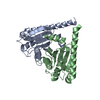
| ||||||||
|---|---|---|---|---|---|---|---|---|---|
| 1 |
| ||||||||
| Unit cell |
|
-Components
| #1: Protein | Mass: 20734.672 Da / Num. of mol.: 2 / Fragment: C-terminal fragment 2 (UNP residues 99 -276 ) Source method: isolated from a genetically manipulated source Source: (gene. exp.) Salmonella typhimurium (bacteria) / Strain: SJW1103 / Gene: motB / Plasmid: PET19b / Production host: #2: Water | ChemComp-HOH / |  Mass: 18.015 Da / Num. of mol.: 469 / Source method: isolated from a natural source / Formula: H2O Mass: 18.015 Da / Num. of mol.: 469 / Source method: isolated from a natural source / Formula: H2O |
|---|
-Experimental details
-Experiment
| Experiment | Method: X-RAY DIFFRACTION / Number of used crystals: 1 |
|---|
- Sample preparation
| Crystal | Density Matthews: 2.17 Å3/Da / Density % sol: 43.42 % |
|---|---|
| Crystal grow | Temperature: 293 K / Method: vapor diffusion, sitting drop / pH: 8.5 Details: 30% PEG 4000, 0.1M Tris-HCl, 0.2M Sodium Acetate, pH 8.5, VAPOR DIFFUSION, SITTING DROP, temperature 293.0K |
-Data collection
| Diffraction | Mean temperature: 35 K |
|---|---|
| Diffraction source | Source: SYNCHROTRON / Site: SPring-8  / Beamline: BL41XU / Wavelength: 1 Å / Beamline: BL41XU / Wavelength: 1 Å |
| Detector | Type: ADSC QUANTUM 315 / Detector: CCD / Date: Feb 22, 2008 |
| Radiation | Monochromator: Double-crystal monochromator / Protocol: SINGLE WAVELENGTH / Monochromatic (M) / Laue (L): M / Scattering type: x-ray |
| Radiation wavelength | Wavelength: 1 Å / Relative weight: 1 |
| Reflection | Resolution: 1.75→35.97 Å / Num. all: 36833 / Num. obs: 36833 / % possible obs: 99.9 % / Observed criterion σ(F): 0 / Observed criterion σ(I): 0 / Redundancy: 5.6 % / Biso Wilson estimate: 16.4 Å2 / Rsym value: 0.01 / Net I/σ(I): 15.1 |
| Reflection shell | Resolution: 1.75→1.84 Å / Redundancy: 5.7 % / Mean I/σ(I) obs: 4.5 / Num. unique all: 5341 / Rsym value: 0.314 / % possible all: 100 |
- Processing
| Software |
| ||||||||||||||||||||||||||||||||||||||||||||||||||||||||||||||||||||||||||||||||
|---|---|---|---|---|---|---|---|---|---|---|---|---|---|---|---|---|---|---|---|---|---|---|---|---|---|---|---|---|---|---|---|---|---|---|---|---|---|---|---|---|---|---|---|---|---|---|---|---|---|---|---|---|---|---|---|---|---|---|---|---|---|---|---|---|---|---|---|---|---|---|---|---|---|---|---|---|---|---|---|---|---|
| Refinement | Method to determine structure: MOLECULAR REPLACEMENT Starting model: PDB ENTRY 2ZOV Resolution: 1.75→35.97 Å / Rfactor Rfree error: 0.005 / Data cutoff high absF: 1558926.39 / Data cutoff low absF: 0 / Isotropic thermal model: RESTRAINED / Cross valid method: THROUGHOUT / σ(F): 0 / Stereochemistry target values: Engh & Huber
| ||||||||||||||||||||||||||||||||||||||||||||||||||||||||||||||||||||||||||||||||
| Solvent computation | Solvent model: FLAT MODEL / Bsol: 44.0914 Å2 / ksol: 0.314714 e/Å3 | ||||||||||||||||||||||||||||||||||||||||||||||||||||||||||||||||||||||||||||||||
| Displacement parameters | Biso mean: 22 Å2
| ||||||||||||||||||||||||||||||||||||||||||||||||||||||||||||||||||||||||||||||||
| Refine analyze |
| ||||||||||||||||||||||||||||||||||||||||||||||||||||||||||||||||||||||||||||||||
| Refinement step | Cycle: LAST / Resolution: 1.75→35.97 Å
| ||||||||||||||||||||||||||||||||||||||||||||||||||||||||||||||||||||||||||||||||
| Refine LS restraints |
| ||||||||||||||||||||||||||||||||||||||||||||||||||||||||||||||||||||||||||||||||
| LS refinement shell | Resolution: 1.75→1.86 Å / Rfactor Rfree error: 0.016 / Total num. of bins used: 6
| ||||||||||||||||||||||||||||||||||||||||||||||||||||||||||||||||||||||||||||||||
| Xplor file |
|
