 Movie
Movie Controller

[English] 日本語
 Yorodumi
- PDB-2z17: Crystal structure of PDZ domain from human Pleckstrin homology, Sec7 -
+ Open data

- Basic information
| Entry | Database: PDB / ID: 2z17 | ||||||
|---|---|---|---|---|---|---|---|
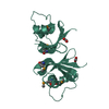
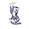
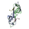
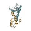
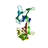
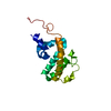
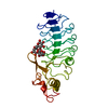
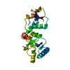
| Title | Crystal structure of PDZ domain from human Pleckstrin homology, Sec7 | ||||||
 Components Components | Pleckstrin homology Sec7 and coiled-coil domains-binding protein | ||||||
 Keywords Keywords | PROTEIN BINDING / PDZ domain / Coiled coil / Cytoplasm / Membrane / Polymorphism / Structural Genomics / NPPSFA / National Project on Protein Structural and Functional Analyses / RIKEN Structural Genomics/Proteomics Initiative / RSGI | ||||||
| Function / homology |  Function and homology information Function and homology informationregulation of cell adhesion / cell cortex / early endosome / nucleoplasm / cytoplasm / cytosol Similarity search - Function | ||||||
| Biological species |  Homo sapiens (human) Homo sapiens (human) | ||||||
| Method |  X-RAY DIFFRACTION / SYNCHROTRON / MAD / Resolution: 2.7 Å X-RAY DIFFRACTION / SYNCHROTRON / MAD / Resolution: 2.7 Å | ||||||
 Authors Authors | Kishishita, S. / Nishino, A. / Murayama, K. / Terada, T. / Shirouzu, M. / Yokoyama, S. / RIKEN Structural Genomics/Proteomics Initiative (RSGI) | ||||||
 Citation Citation | Journal: To be Published Title: Crystal structure of PDZ domain from human Pleckstrin homology, Sec7 Authors: Kishishita, S. / Nishino, A. / Murayama, K. / Terada, T. / Shirouzu, M. / Yokoyama, S. | ||||||
| History |
|
- Structure visualization
| Structure viewer | Molecule: MolmilJmol/JSmol |
|---|
- Downloads & links
-Download
| PDBx/mmCIF format | 2z17.cif.gz | 31 KB | Display | PDBx/mmCIF format |
|---|---|---|---|---|
| PDB format | pdb2z17.ent.gz | 20.6 KB | Display | PDB format |
| PDBx/mmJSON format | 2z17.json.gz | Tree view | PDBx/mmJSON format | |
| Others |  Other downloads Other downloads |
-Validation report
| Arichive directory | https://data.pdbj.org/pub/pdb/validation_reports/z1/2z17ftp://data.pdbj.org/pub/pdb/validation_reports/z1/2z17 | HTTPS FTP |
|---|
-Related structure data
| Similar structure data | |
|---|---|
| Other databases |


-Links
 PDBj
PDBj- Assembly
| Deposited unit | 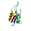
| ||||||||
|---|---|---|---|---|---|---|---|---|---|
| 1 | 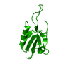
| ||||||||
| Unit cell |
| ||||||||
| Components on special symmetry positions |
|
-Components
| #1: Protein | Mass: 11304.448 Da / Num. of mol.: 1 / Fragment: PDZ domain Source method: isolated from a genetically manipulated source Source: (gene. exp.) Homo sapiens (human) / Plasmid: PK060110-70 / Production host: Cell-free protein synthesis / References: UniProt: O60759 |
|---|---|
| #2: Water | ChemComp-HOH /  Mass: 18.015 Da / Num. of mol.: 11 / Source method: isolated from a natural source / Formula: H2O Mass: 18.015 Da / Num. of mol.: 11 / Source method: isolated from a natural source / Formula: H2O |
-Experimental details
-Experiment
| Experiment | Method: X-RAY DIFFRACTION / Number of used crystals: 1 |
|---|
- Sample preparation
| Crystal | Density Matthews: 2.25 Å3/Da / Density % sol: 45.32 % |
|---|---|
| Crystal grow | Temperature: 293 K / Method: vapor diffusion, hanging drop / pH: 7.5 Details: 0.1M HEPES-Na, 2.0M Ammonium Sulfate, pH 7.5, VAPOR DIFFUSION, HANGING DROP, temperature 293K |
-Data collection
| Diffraction | Mean temperature: 100 K | ||||||||||||
|---|---|---|---|---|---|---|---|---|---|---|---|---|---|
| Diffraction source | Source: SYNCHROTRON / Site: Photon Factory  / Beamline: BL-5A / Wavelength: 0.9789, 0.9794, 0.964 / Beamline: BL-5A / Wavelength: 0.9789, 0.9794, 0.964 | ||||||||||||
| Detector | Type: ADSC QUANTUM 315 / Detector: CCD / Date: May 11, 2006 / Details: mirrors | ||||||||||||
| Radiation | Monochromator: Si II / Protocol: MAD / Monochromatic (M) / Laue (L): M / Scattering type: x-ray | ||||||||||||
| Radiation wavelength |
| ||||||||||||
| Reflection | Resolution: 2.5→50 Å / Num. obs: 3931 / % possible obs: 99.7 % / Observed criterion σ(I): -3 / Redundancy: 19.5 % / Biso Wilson estimate: 38.2 Å2 / Rsym value: 0.061 / Net I/σ(I): 41.9 | ||||||||||||
| Reflection shell | Resolution: 2.5→2.63 Å / Mean I/σ(I) obs: 15.6 / Rsym value: 0.232 / % possible all: 99.6 |
- Processing
| Software |
| ||||||||||||||||||||||||||||||||||||||||||||||||||||||||||||
|---|---|---|---|---|---|---|---|---|---|---|---|---|---|---|---|---|---|---|---|---|---|---|---|---|---|---|---|---|---|---|---|---|---|---|---|---|---|---|---|---|---|---|---|---|---|---|---|---|---|---|---|---|---|---|---|---|---|---|---|---|---|
| Refinement | Method to determine structure: MAD / Resolution: 2.7→28.54 Å / Rfactor Rfree error: 0.015 / Data cutoff high absF: 145252.75 / Data cutoff low absF: 0 / Isotropic thermal model: RESTRAINED / Cross valid method: THROUGHOUT / σ(F): 0
| ||||||||||||||||||||||||||||||||||||||||||||||||||||||||||||
| Solvent computation | Solvent model: FLAT MODEL / Bsol: 46.1437 Å2 / ksol: 0.340996 e/Å3 | ||||||||||||||||||||||||||||||||||||||||||||||||||||||||||||
| Displacement parameters | Biso mean: 44 Å2
| ||||||||||||||||||||||||||||||||||||||||||||||||||||||||||||
| Refine analyze |
| ||||||||||||||||||||||||||||||||||||||||||||||||||||||||||||
| Refinement step | Cycle: LAST / Resolution: 2.7→28.54 Å
| ||||||||||||||||||||||||||||||||||||||||||||||||||||||||||||
| Refine LS restraints |
| ||||||||||||||||||||||||||||||||||||||||||||||||||||||||||||
| LS refinement shell | Resolution: 2.7→2.87 Å / Rfactor Rfree error: 0.07 / Total num. of bins used: 6
| ||||||||||||||||||||||||||||||||||||||||||||||||||||||||||||
| Xplor file |
|
