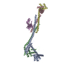
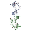
[English] 日本語
Yorodumi
- PDB-2yq3: Structure of BVDV1 envelope glycoprotein E2, pH5 -

+
Open data


ID or keywords:

Loading...

-
Basic information

Entry
Database: PDB / ID: 2yq3
TitleStructure of BVDV1 envelope glycoprotein E2, pH5
ComponentsBVDV1 E2
KeywordsVIRAL PROTEIN / PESTIVIRUS / VIRUS FUSION
Function / homology
Function and homology information


pestivirus NS3 polyprotein peptidase / ribonuclease T2 / serine-type exopeptidase activity / ribonuclease T2 activity / : / nucleoside-triphosphate phosphatase / protein complex oligomerization / monoatomic ion channel activity / symbiont-mediated suppression of host cytoplasmic pattern recognition receptor signaling pathway via inhibition of IRF3 activity / clathrin-dependent endocytosis of virus by host cell ...pestivirus NS3 polyprotein peptidase / ribonuclease T2 / serine-type exopeptidase activity / ribonuclease T2 activity / : / nucleoside-triphosphate phosphatase / protein complex oligomerization / monoatomic ion channel activity / symbiont-mediated suppression of host cytoplasmic pattern recognition receptor signaling pathway via inhibition of IRF3 activity / clathrin-dependent endocytosis of virus by host cell / Hydrolases; Acting on peptide bonds (peptidases); Cysteine endopeptidases / RNA helicase activity / host cell endoplasmic reticulum membrane / viral protein processing / RNA helicase / induction by virus of host autophagy / RNA-directed RNA polymerase / viral RNA genome replication / cysteine-type endopeptidase activity / RNA-dependent RNA polymerase activity / serine-type endopeptidase activity / fusion of virus membrane with host endosome membrane / virion attachment to host cell / GTP binding / virion membrane / ATP hydrolysis activity / proteolysis / RNA binding / ATP binding / membrane
Similarity search - Function
Alpha-Beta Plaits - #50 / Pestivirus envelope glycoprotein E2, domain B / Alpha-Beta Plaits / Pestivirus envelope glycoprotein E2, domain A / Pestivirus envelope glycoprotein E2, domain B / Capsid protein C, pestivirus / Pestivirus nonstructural protein 2 / Pestivirus NS2, peptidase C74 / Capsid protein C, pestivirus / Pestivirus NS2 peptidase ...Alpha-Beta Plaits - #50 / Pestivirus envelope glycoprotein E2, domain B / Alpha-Beta Plaits / Pestivirus envelope glycoprotein E2, domain A / Pestivirus envelope glycoprotein E2, domain B / Capsid protein C, pestivirus / Pestivirus nonstructural protein 2 / Pestivirus NS2, peptidase C74 / Capsid protein C, pestivirus / Pestivirus NS2 peptidase / Pestivirus nonstructural protein 2 (NS2) protease domain profile. / Pestivirus NS3, peptidase S31 / Pestivirus envelope glycoprotein E2 / Pestivirus envelope glycoprotein E2, domain D / Pestivirus NS3 polyprotein peptidase S31 / Pestivirus envelope glycoprotein E2 / Pestivirus NS3 protease (NS3pro) domain profile. / Peptidase C53, pestivirus Npro, interaction domain / Peptidase C53, pestivirus Npro / Pestivirus Npro endopeptidase C53 / Pestivirus N-terminal protease Npro domain profile. / Ribonuclease T2, His active site 2 / Ribonuclease T2 family histidine active site 2. / Ribonuclease T2-like superfamily / Other non-globular / RNA dependent RNA polymerase, hepatitis C virus / Viral RNA dependent RNA polymerase / : / Flavivirus NS3 helicase, C-terminal helical domain / DEAD box, Flavivirus / Flavivirus DEAD domain / Helicase conserved C-terminal domain / Special / helicase superfamily c-terminal domain / Superfamilies 1 and 2 helicase C-terminal domain profile. / Superfamilies 1 and 2 helicase ATP-binding type-1 domain profile. / DEAD-like helicases superfamily / Helicase, C-terminal / Helicase superfamily 1/2, ATP-binding domain / Reverse transcriptase/Diguanylate cyclase domain / RNA-directed RNA polymerase, catalytic domain / RdRp of positive ssRNA viruses catalytic domain profile. / DNA/RNA polymerase superfamily / Immunoglobulin-like / Sandwich / P-loop containing nucleoside triphosphate hydrolase / Mainly Beta
Similarity search - Domain/homology
Biological speciesBOVINE VIRAL DIARRHEA VIRUS 1
MethodX-RAY DIFFRACTION / SYNCHROTRON / MOLECULAR REPLACEMENT / Resolution: 3.29 Å
AuthorsEl Omari, K. / Iourin, O. / Harlos, K. / Grimes, J.M. / Stuart, D.I.
CitationJournal: Cell Rep. / Year: 2013
Title: Structure of a Pestivirus Envelope Glycoprotein E2 Clarifies its Role in Cell Entry.
Authors: El Omari, K. / Iourin, O. / Harlos, K. / Grimes, J.M. / Stuart, D.I.
History
DepositionNov 4, 2012Deposition site: PDBE / Processing site: PDBE
Revision 1.0Jan 16, 2013Provider: repository / Type: Initial release
Revision 1.1Feb 20, 2013Group: Database references
Revision 1.2Nov 26, 2014Group: Other
Revision 1.3Apr 3, 2019Group: Data collection / Derived calculations ...Data collection / Derived calculations / Other / Source and taxonomy
Category: entity_src_gen / pdbx_database_proc ...entity_src_gen / pdbx_database_proc / pdbx_database_status / struct_conn
Item: _entity_src_gen.pdbx_host_org_cell_line / _pdbx_database_status.recvd_author_approval / _struct_conn.pdbx_leaving_atom_flag
Revision 1.4Jul 29, 2020Group: Data collection / Derived calculations ...Data collection / Derived calculations / Other / Structure summary
Category: chem_comp / entity ...chem_comp / entity / pdbx_chem_comp_identifier / pdbx_database_status / pdbx_entity_nonpoly / struct_conn / struct_site / struct_site_gen
Item: _chem_comp.name / _chem_comp.type ..._chem_comp.name / _chem_comp.type / _entity.pdbx_description / _pdbx_database_status.status_code_sf / _pdbx_entity_nonpoly.name / _struct_conn.pdbx_role
Description: Carbohydrate remediation / Provider: repository / Type: Remediation
Revision 1.5Dec 20, 2023Group: Data collection / Database references ...Data collection / Database references / Refinement description / Structure summary
Category: chem_comp / chem_comp_atom ...chem_comp / chem_comp_atom / chem_comp_bond / database_2 / pdbx_initial_refinement_model
Item: _chem_comp.pdbx_synonyms / _database_2.pdbx_DOI / _database_2.pdbx_database_accession

-
Structure visualization

Structure viewerMolecule:
MolmilJmol/JSmol

Downloads & links

-
Assembly

Deposited unit
A: BVDV1 E2
B: BVDV1 E2
hetero molecules


Theoretical massNumber of molelcules
Total (without water)77,93510
Polymers76,1652
Non-polymers1,7708
Water0
1


  • Idetical with deposited unit
  • defined by author&software
TypeNameSymmetry operationNumber
identity operation1_555x,y,z1
Buried area6720 Å2
ΔGint-9.6 kcal/mol
Surface area31430 Å2
MethodPISA
Unit cell
Length a, b, c (Å)160.736, 49.695, 136.422
Angle α, β, γ (deg.)90.00, 90.00, 90.00
Int Tables number18
Space group name H-MP21212

-
Components

#1: Protein BVDV1 E2


Mass: 38082.441 Da / Num. of mol.: 2 / Fragment: BVDV1 E2 ECTODOMAIN, RESIDUES 1-337 / Mutation: YES
Source method: isolated from a genetically manipulated source
Source: (gene. exp.) BOVINE VIRAL DIARRHEA VIRUS 1 / Strain: PE515 / Plasmid: PHLSEC / Cell line (production host): HEK293T / Production host: HOMO SAPIENS (human) / References: UniProt: Q01499*PLUS
#2: Sugar
ChemComp-NAG / 2-acetamido-2-deoxy-beta-D-glucopyranose / N-acetyl-beta-D-glucosamine / 2-acetamido-2-deoxy-beta-D-glucose / 2-acetamido-2-deoxy-D-glucose / 2-acetamido-2-deoxy-glucose / N-ACETYL-D-GLUCOSAMINE / N-Acetylglucosamine


Type: D-saccharide, beta linking / Mass: 221.208 Da / Num. of mol.: 8
Source method: isolated from a genetically manipulated source
Formula: C8H15NO6
IdentifierTypeProgram
DGlcpNAcbCONDENSED IUPAC CARBOHYDRATE SYMBOLGMML 1.0
N-acetyl-b-D-glucopyranosamineCOMMON NAMEGMML 1.0
b-D-GlcpNAcIUPAC CARBOHYDRATE SYMBOLPDB-CARE 1.0
GlcNAcSNFG CARBOHYDRATE SYMBOLGMML 1.0
Sequence detailsMUTATION: Y9S, G94E

-
Experimental details

-
Experiment

ExperimentMethod: X-RAY DIFFRACTION / Number of used crystals: 1

-
Sample preparation

CrystalDensity Matthews: 3.36 Å3/Da / Density % sol: 63.4 % / Description: NONE
Crystal growpH: 5
Details: 16% PEG 6000, 79 MM CITRIC BUFFER PH 5.0, 20MM MGSO4

-
Data collection

DiffractionMean temperature: 100 K
Diffraction sourceSource: SYNCHROTRON / Site: Diamond / Beamline: I24 / Wavelength: 1.0341
DetectorType: DECTRIS PILATUS 6M / Detector: PIXEL
RadiationProtocol: SINGLE WAVELENGTH / Monochromatic (M) / Laue (L): M / Scattering type: x-ray
Radiation wavelengthWavelength: 1.0341 Å / Relative weight: 1
ReflectionResolution: 3.3→50 Å / Num. obs: 17239 / % possible obs: 99.9 % / Observed criterion σ(I): 1.5 / Redundancy: 4.9 % / Biso Wilson estimate: 75.46 Å2 / Rmerge(I) obs: 0.24 / Net I/σ(I): 6.1
Reflection shellResolution: 3.3→3.42 Å / Redundancy: 4.8 % / Rmerge(I) obs: 0.92 / Mean I/σ(I) obs: 1.5 / % possible all: 99.8

-
Processing

Software
NameVersionClassification
BUSTER2.11.2refinement
HKL-2000data reduction
SCALEPACKdata scaling
MOLREPphasing
RefinementMethod to determine structure: MOLECULAR REPLACEMENT
Starting model: PDB ENTRY 2YQ2
Resolution: 3.29→47.48 Å / Cor.coef. Fo:Fc: 0.8241 / Cor.coef. Fo:Fc free: 0.821 / SU R Cruickshank DPI: 2.592 / Cross valid method: THROUGHOUT / σ(F): 0 / SU R Blow DPI: 4.522 / SU Rfree Blow DPI: 0.424 / SU Rfree Cruickshank DPI: 0.427
RfactorNum. reflection% reflectionSelection details
Rfree0.2637 873 5.07 %RANDOM
Rwork0.2526 ---
obs0.2531 17209 99.29 %-
Displacement parametersBiso mean: 82.96 Å2
Baniso -1Baniso -2Baniso -3
1--15.1136 Å20 Å20 Å2
2--7.7565 Å20 Å2
3---7.3571 Å2
Refine analyzeLuzzati coordinate error obs: 0.885 Å
Refinement stepCycle: LAST / Resolution: 3.29→47.48 Å
ProteinNucleic acidLigandSolventTotal
Num. atoms4083 0 112 0 4195
Refine LS restraints
Refine-IDTypeDev idealNumberRestraint functionWeight
X-RAY DIFFRACTIONt_bond_d0.0074304HARMONIC2
X-RAY DIFFRACTIONt_angle_deg0.995836HARMONIC2
X-RAY DIFFRACTIONt_dihedral_angle_d1979SINUSOIDAL2
X-RAY DIFFRACTIONt_incorr_chiral_ct
X-RAY DIFFRACTIONt_pseud_angle
X-RAY DIFFRACTIONt_trig_c_planes102HARMONIC2
X-RAY DIFFRACTIONt_gen_planes616HARMONIC5
X-RAY DIFFRACTIONt_it4304HARMONIC20
X-RAY DIFFRACTIONt_nbd
X-RAY DIFFRACTIONt_omega_torsion2.23
X-RAY DIFFRACTIONt_other_torsion3.11
X-RAY DIFFRACTIONt_improper_torsion
X-RAY DIFFRACTIONt_chiral_improper_torsion582SEMIHARMONIC5
X-RAY DIFFRACTIONt_sum_occupancies
X-RAY DIFFRACTIONt_utility_distance
X-RAY DIFFRACTIONt_utility_angle
X-RAY DIFFRACTIONt_utility_torsion
X-RAY DIFFRACTIONt_ideal_dist_contact4298SEMIHARMONIC4
LS refinement shellResolution: 3.29→3.49 Å / Total num. of bins used: 9
RfactorNum. reflection% reflection
Rfree0.206 148 5.58 %
Rwork0.2395 2506 -
all0.2377 2654 -
obs--99.29 %
Refinement TLS params.

Method: refined / Refine-ID: X-RAY DIFFRACTION

IDL112)L122)L132)L222)L232)L332)S11 (Å °)S12 (Å °)S13 (Å °)S21 (Å °)S22 (Å °)S23 (Å °)S31 (Å °)S32 (Å °)S33 (Å °)T112)T122)T132)T222)T232)T332)Origin x (Å)Origin y (Å)Origin z (Å)
10.24650.23140.00170.94471.86885.3911-0.03140.01510.0755-0.27420.0008-0.0970.07-0.28830.03060.11220.04730.0167-0.05320.0512-0.0062-50.72378.9575-27.1014
20.70150.2015-1.21510.0245-0.25983.6253-0.06870.0150.19130.24090.0931-0.19020.1947-0.2224-0.02440.1896-0.01050.0041-0.1150.0083-0.0182-33.7222.9117-104.166
Refinement TLS group
IDRefine-IDRefine TLS-IDSelection details
1X-RAY DIFFRACTION1CHAIN A
2X-RAY DIFFRACTION2CHAIN B

+
About Yorodumi

-
News

-
Feb 9, 2022. New format data for meta-information of EMDB entries

New format data for meta-information of EMDB entries

  • Version 3 of the EMDB header file is now the official format.
  • The previous official version 1.9 will be removed from the archive.

Related info.:EMDB header

External links:wwPDB to switch to version 3 of the EMDB data model

-
Aug 12, 2020. Covid-19 info

Covid-19 info

URL: https://pdbj.org/emnavi/covid19.php

New page: Covid-19 featured information page in EM Navigator.

Related info.:Covid-19 info / Mar 5, 2020. Novel coronavirus structure data

+
Mar 5, 2020. Novel coronavirus structure data

Novel coronavirus structure data

Related info.:Yorodumi Speices / Aug 12, 2020. Covid-19 info

External links:COVID-19 featured content - PDBj / Molecule of the Month (242):Coronavirus Proteases

+
Jan 31, 2019. EMDB accession codes are about to change! (news from PDBe EMDB page)

EMDB accession codes are about to change! (news from PDBe EMDB page)

  • The allocation of 4 digits for EMDB accession codes will soon come to an end. Whilst these codes will remain in use, new EMDB accession codes will include an additional digit and will expand incrementally as the available range of codes is exhausted. The current 4-digit format prefixed with “EMD-” (i.e. EMD-XXXX) will advance to a 5-digit format (i.e. EMD-XXXXX), and so on. It is currently estimated that the 4-digit codes will be depleted around Spring 2019, at which point the 5-digit format will come into force.
  • The EM Navigator/Yorodumi systems omit the EMD- prefix.

Related info.:Q: What is EMD? / ID/Accession-code notation in Yorodumi/EM Navigator

External links:EMDB Accession Codes are Changing Soon! / Contact to PDBj

+
Jul 12, 2017. Major update of PDB

Major update of PDB

  • wwPDB released updated PDB data conforming to the new PDBx/mmCIF dictionary.
  • This is a major update changing the version number from 4 to 5, and with Remediation, in which all the entries are updated.
  • In this update, many items about electron microscopy experimental information are reorganized (e.g. em_software).
  • Now, EM Navigator and Yorodumi are based on the updated data.

External links:wwPDB Remediation / Enriched Model Files Conforming to OneDep Data Standards Now Available in the PDB FTP Archive

-
Yorodumi

Thousand views of thousand structures

  • Yorodumi is a browser for structure data from EMDB, PDB, SASBDB, etc.
  • This page is also the successor to EM Navigator detail page, and also detail information page/front-end page for Omokage search.
  • The word "yorodu" (or yorozu) is an old Japanese word meaning "ten thousand". "mi" (miru) is to see.

Related info.:EMDB / PDB / SASBDB / Comparison of 3 databanks / Yorodumi Search / Aug 31, 2016. New EM Navigator & Yorodumi / Yorodumi Papers / Jmol/JSmol / Function and homology information / Changes in new EM Navigator and Yorodumi

Read more