 Movie
Movie Controller


+ Open data

- Basic information
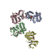
| Entry | Database: PDB / ID: 6kbi | |||||||||
|---|---|---|---|---|---|---|---|---|---|---|
| Title | Crystal structure of ErbB3 N418Q mutant | |||||||||
 Components Components | Receptor tyrosine-protein kinase erbB-3 | |||||||||
 Keywords Keywords | SIGNALING PROTEIN / Receptor protein / tyrosine kinase / ErbB-3 | |||||||||
| Function / homology |  Function and homology information Function and homology informationpositive regulation of cardiac muscle tissue development / neuregulin binding / cranial nerve development / Schwann cell differentiation / neuregulin receptor activity / negative regulation of secretion / endocardial cushion development / ERBB3:ERBB2 complex / GRB7 events in ERBB2 signaling / positive regulation of calcineurin-NFAT signaling cascade ...positive regulation of cardiac muscle tissue development / neuregulin binding / cranial nerve development / Schwann cell differentiation / neuregulin receptor activity / negative regulation of secretion / endocardial cushion development / ERBB3:ERBB2 complex / GRB7 events in ERBB2 signaling / positive regulation of calcineurin-NFAT signaling cascade / peripheral nervous system development / negative regulation of cell adhesion / negative regulation of motor neuron apoptotic process / ErbB-3 class receptor binding / motor neuron apoptotic process / ERBB2 Activates PTK6 Signaling / growth factor binding / ERBB2-ERBB3 signaling pathway / ERBB2 Regulates Cell Motility / protein tyrosine kinase activator activity / Signaling by ERBB4 / PI3K events in ERBB2 signaling / lateral plasma membrane / negative regulation of signal transduction / Schwann cell development / extrinsic apoptotic signaling pathway in absence of ligand / Signaling by ERBB2 / myelination / SHC1 events in ERBB2 signaling / cell surface receptor protein tyrosine kinase signaling pathway / basal plasma membrane / positive regulation of epithelial cell proliferation / Downregulation of ERBB2:ERBB3 signaling / wound healing / phosphatidylinositol 3-kinase/protein kinase B signal transduction / Signaling by ERBB2 TMD/JMD mutants / receptor protein-tyrosine kinase / Signaling by ERBB2 KD Mutants / epidermal growth factor receptor signaling pathway / Downregulation of ERBB2 signaling / Constitutive Signaling by Aberrant PI3K in Cancer / neuron differentiation / transmembrane signaling receptor activity / PIP3 activates AKT signaling / regulation of cell population proliferation / heart development / PI5P, PP2A and IER3 Regulate PI3K/AKT Signaling / RAF/MAP kinase cascade / neuron apoptotic process / basolateral plasma membrane / negative regulation of neuron apoptotic process / protein kinase activity / positive regulation of MAPK cascade / positive regulation of phosphatidylinositol 3-kinase/protein kinase B signal transduction / signaling receptor complex / apical plasma membrane / protein heterodimerization activity / ubiquitin protein ligase binding / positive regulation of gene expression / negative regulation of apoptotic process / signal transduction / : / ATP binding / identical protein binding / plasma membrane Similarity search - Function | |||||||||
| Biological species |  Homo sapiens (human) Homo sapiens (human) | |||||||||
| Method |  X-RAY DIFFRACTION / SYNCHROTRON / MOLECULAR REPLACEMENT / Resolution: 3 Å X-RAY DIFFRACTION / SYNCHROTRON / MOLECULAR REPLACEMENT / Resolution: 3 Å | |||||||||
 Authors Authors | Kato, K. / Yao, M. | |||||||||
 Citation Citation | Journal: To Be Published Title: Crystal structure of ErbB3 N418Q mutant Authors: Takahashi, M. / Kato, K. / Yao, M. | |||||||||
| History |
|
- Structure visualization
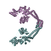
| Structure viewer | Molecule: MolmilJmol/JSmol |
|---|
- Downloads & links
-Download
| PDBx/mmCIF format | 6kbi.cif.gz | 233.7 KB | Display | PDBx/mmCIF format |
|---|---|---|---|---|
| PDB format | pdb6kbi.ent.gz | 187.2 KB | Display | PDB format |
| PDBx/mmJSON format | 6kbi.json.gz | Tree view | PDBx/mmJSON format | |
| Others |  Other downloads Other downloads |
-Validation report
| Arichive directory | https://data.pdbj.org/pub/pdb/validation_reports/kb/6kbiftp://data.pdbj.org/pub/pdb/validation_reports/kb/6kbi | HTTPS FTP |
|---|
-Related structure data
| Related structure data | 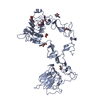 1m6bS S: Starting model for refinement |
|---|---|
| Similar structure data |
-Links
 PDBj
PDBj


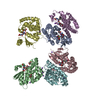
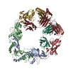
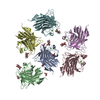
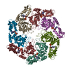
- Assembly
| Deposited unit | 
| ||||||||
|---|---|---|---|---|---|---|---|---|---|
| 1 | 
| ||||||||
| Unit cell |
|
-Components
| #1: Protein | Mass: 68303.500 Da / Num. of mol.: 2 / Mutation: N418Q Source method: isolated from a genetically manipulated source Source: (gene. exp.) Homo sapiens (human) / Gene: ERBB3, HER3 / Production host:   Cricetulus griseus (Chinese hamster) Cricetulus griseus (Chinese hamster)References: UniProt: P21860, receptor protein-tyrosine kinase #2: Polysaccharide | Source method: isolated from a genetically manipulated source #3: Sugar | ChemComp-NAG /   Type: D-saccharide, beta linking / Mass: 221.208 Da / Num. of mol.: 13 Type: D-saccharide, beta linking / Mass: 221.208 Da / Num. of mol.: 13Source method: isolated from a genetically manipulated source Formula: C8H15NO6 Has ligand of interest | N | Has protein modification | Y | |
|---|
-Experimental details
-Experiment
| Experiment | Method: X-RAY DIFFRACTION / Number of used crystals: 1 |
|---|
- Sample preparation
| Crystal | Density Matthews: 3.4 Å3/Da / Density % sol: 63.81 % |
|---|---|
| Crystal grow | Temperature: 293 K / Method: vapor diffusion, hanging drop / pH: 8.5 Details: 0.2 M lithium sulfate, 0.1 M Tris buffer (pH 8.5) and 11% (w/v) PEG4000. |
-Data collection
| Diffraction | Mean temperature: 100 K / Serial crystal experiment: N |
|---|---|
| Diffraction source | Source: SYNCHROTRON / Site: SPring-8  / Beamline: BL38B1 / Wavelength: 1 Å / Beamline: BL38B1 / Wavelength: 1 Å |
| Detector | Type: ADSC QUANTUM 315r / Detector: CCD / Date: May 11, 2016 |
| Radiation | Protocol: SINGLE WAVELENGTH / Monochromatic (M) / Laue (L): M / Scattering type: x-ray |
| Radiation wavelength | Wavelength: 1 Å / Relative weight: 1 |
| Reflection | Resolution: 3→50 Å / Num. obs: 36623 / % possible obs: 99.1 % / Redundancy: 3.7 % / Rrim(I) all: 0.105 / Net I/σ(I): 11.4 |
| Reflection shell | Resolution: 3→3.18 Å / Redundancy: 3.7 % / Mean I/σ(I) obs: 2.2 / Num. unique obs: 5774 / Rrim(I) all: 0.564 / % possible all: 97.9 |
- Processing
| Software |
| ||||||||||||||||||||||||||||||||||||||||||||||||||||||||||||||||||||||||||||||||||||||||||||||||||
|---|---|---|---|---|---|---|---|---|---|---|---|---|---|---|---|---|---|---|---|---|---|---|---|---|---|---|---|---|---|---|---|---|---|---|---|---|---|---|---|---|---|---|---|---|---|---|---|---|---|---|---|---|---|---|---|---|---|---|---|---|---|---|---|---|---|---|---|---|---|---|---|---|---|---|---|---|---|---|---|---|---|---|---|---|---|---|---|---|---|---|---|---|---|---|---|---|---|---|---|
| Refinement | Method to determine structure: MOLECULAR REPLACEMENT Starting model: 1M6B Resolution: 3→46.623 Å / SU ML: 0.44 / Cross valid method: FREE R-VALUE / σ(F): 1.35 / Phase error: 32
| ||||||||||||||||||||||||||||||||||||||||||||||||||||||||||||||||||||||||||||||||||||||||||||||||||
| Solvent computation | Shrinkage radii: 0.9 Å / VDW probe radii: 1.11 Å | ||||||||||||||||||||||||||||||||||||||||||||||||||||||||||||||||||||||||||||||||||||||||||||||||||
| Refinement step | Cycle: LAST / Resolution: 3→46.623 Å
| ||||||||||||||||||||||||||||||||||||||||||||||||||||||||||||||||||||||||||||||||||||||||||||||||||
| Refine LS restraints |
| ||||||||||||||||||||||||||||||||||||||||||||||||||||||||||||||||||||||||||||||||||||||||||||||||||
| LS refinement shell |
|
