 Movie
Movie Controller


+ Open data

- Basic information
| Entry | Database: PDB / ID: 2y5b | ||||||
|---|---|---|---|---|---|---|---|
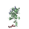
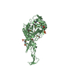
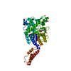
| Title | Structure of USP21 in complex with linear diubiquitin-aldehyde | ||||||
 Components Components |
| ||||||
 Keywords Keywords | PROTEIN BINDING/HYDROLASE / PROTEIN BINDING-HYDROLASE COMPLEX / UBIQUITIN / UBIQUITIN SPECIFIC PROTEASE / USP / NEDD8 / ISG15 / CELL SIGNALING | ||||||
| Function / homology |  Function and homology information Function and homology informationdeNEDDylase activity / hypothalamus gonadotrophin-releasing hormone neuron development / female meiosis I / positive regulation of protein monoubiquitination / TNFR1-induced proapoptotic signaling / fat pad development / mitochondrion transport along microtubule / female gonad development / seminiferous tubule development / male meiosis I ...deNEDDylase activity / hypothalamus gonadotrophin-releasing hormone neuron development / female meiosis I / positive regulation of protein monoubiquitination / TNFR1-induced proapoptotic signaling / fat pad development / mitochondrion transport along microtubule / female gonad development / seminiferous tubule development / male meiosis I / protein deubiquitination / positive regulation of intrinsic apoptotic signaling pathway by p53 class mediator / energy homeostasis / neuron projection morphogenesis / regulation of proteasomal protein catabolic process / Maturation of protein E / Maturation of protein E / cysteine-type peptidase activity / ER Quality Control Compartment (ERQC) / Myoclonic epilepsy of Lafora / FLT3 signaling by CBL mutants / Constitutive Signaling by NOTCH1 HD Domain Mutants / IRAK2 mediated activation of TAK1 complex / Prevention of phagosomal-lysosomal fusion / Alpha-protein kinase 1 signaling pathway / Glycogen synthesis / IRAK1 recruits IKK complex / IRAK1 recruits IKK complex upon TLR7/8 or 9 stimulation / Endosomal Sorting Complex Required For Transport (ESCRT) / Membrane binding and targetting of GAG proteins / Negative regulation of FLT3 / Regulation of TBK1, IKKε (IKBKE)-mediated activation of IRF3, IRF7 / PTK6 Regulates RTKs and Their Effectors AKT1 and DOK1 / transcription initiation-coupled chromatin remodeling / Regulation of TBK1, IKKε-mediated activation of IRF3, IRF7 upon TLR3 ligation / IRAK2 mediated activation of TAK1 complex upon TLR7/8 or 9 stimulation / NOTCH2 Activation and Transmission of Signal to the Nucleus / TICAM1,TRAF6-dependent induction of TAK1 complex / TICAM1-dependent activation of IRF3/IRF7 / APC/C:Cdc20 mediated degradation of Cyclin B / Regulation of FZD by ubiquitination / Downregulation of ERBB4 signaling / APC-Cdc20 mediated degradation of Nek2A / p75NTR recruits signalling complexes / InlA-mediated entry of Listeria monocytogenes into host cells / TRAF6 mediated IRF7 activation in TLR7/8 or 9 signaling / Regulation of pyruvate metabolism / NF-kB is activated and signals survival / TRAF6-mediated induction of TAK1 complex within TLR4 complex / regulation of neuron apoptotic process / Downregulation of ERBB2:ERBB3 signaling / Pexophagy / Regulation of innate immune responses to cytosolic DNA / NRIF signals cell death from the nucleus / Regulation of PTEN localization / Activated NOTCH1 Transmits Signal to the Nucleus / VLDLR internalisation and degradation / positive regulation of protein ubiquitination / Synthesis of active ubiquitin: roles of E1 and E2 enzymes / TICAM1, RIP1-mediated IKK complex recruitment / Regulation of BACH1 activity / Translesion synthesis by REV1 / MAP3K8 (TPL2)-dependent MAPK1/3 activation / Degradation of CDH1 / Translesion synthesis by POLK / InlB-mediated entry of Listeria monocytogenes into host cell / JNK (c-Jun kinases) phosphorylation and activation mediated by activated human TAK1 / Activation of IRF3, IRF7 mediated by TBK1, IKKε (IKBKE) / Josephin domain DUBs / Downregulation of TGF-beta receptor signaling / Translesion synthesis by POLI / Gap-filling DNA repair synthesis and ligation in GG-NER / IKK complex recruitment mediated by RIP1 / Degradation of CRY and PER proteins / Regulation of activated PAK-2p34 by proteasome mediated degradation / PINK1-PRKN Mediated Mitophagy / regulation of mitochondrial membrane potential / TGF-beta receptor signaling in EMT (epithelial to mesenchymal transition) / TNFR1-induced NF-kappa-B signaling pathway / Autodegradation of Cdh1 by Cdh1:APC/C / TCF dependent signaling in response to WNT / Regulation of NF-kappa B signaling / APC/C:Cdc20 mediated degradation of Securin / N-glycan trimming in the ER and Calnexin/Calreticulin cycle / activated TAK1 mediates p38 MAPK activation / Asymmetric localization of PCP proteins / Ubiquitin-dependent degradation of Cyclin D / SCF-beta-TrCP mediated degradation of Emi1 / NIK-->noncanonical NF-kB signaling / Regulation of signaling by CBL / TNFR2 non-canonical NF-kB pathway / AUF1 (hnRNP D0) binds and destabilizes mRNA / Negative regulators of DDX58/IFIH1 signaling / NOTCH3 Activation and Transmission of Signal to the Nucleus / Assembly of the pre-replicative complex / Negative regulation of FGFR3 signaling / Fanconi Anemia Pathway / Peroxisomal protein import / Vpu mediated degradation of CD4 / Deactivation of the beta-catenin transactivating complex Similarity search - Function | ||||||
| Biological species |  HOMO SAPIENS (human) HOMO SAPIENS (human) | ||||||
| Method |  X-RAY DIFFRACTION / SYNCHROTRON / MOLECULAR REPLACEMENT / Resolution: 2.7 Å X-RAY DIFFRACTION / SYNCHROTRON / MOLECULAR REPLACEMENT / Resolution: 2.7 Å | ||||||
 Authors Authors | Ye, Y. / Akutsu, M. / Reyes-Turcu, F. / Enchev, R.I. / Wilkinson, K.D. / Komander, D. | ||||||
 Citation Citation | Journal: Embo Rep. / Year: 2011 Title: Polyubiquitin Binding and Cross-Reactivity in the Usp Domain Deubiquitinase Usp21. Authors: Ye, Y. / Akutsu, M. / Reyes-Turcu, F. / Enchev, R.I. / Wilkinson, K.D. / Komander, D. | ||||||
| History |
|
- Structure visualization
| Structure viewer | Molecule: MolmilJmol/JSmol |
|---|
- Downloads & links
-Download
| PDBx/mmCIF format | 2y5b.cif.gz | 191.8 KB | Display | PDBx/mmCIF format |
|---|---|---|---|---|
| PDB format | pdb2y5b.ent.gz | 148 KB | Display | PDB format |
| PDBx/mmJSON format | 2y5b.json.gz | Tree view | PDBx/mmJSON format | |
| Others |  Other downloads Other downloads |
-Validation report
| Arichive directory | https://data.pdbj.org/pub/pdb/validation_reports/y5/2y5bftp://data.pdbj.org/pub/pdb/validation_reports/y5/2y5b | HTTPS FTP |
|---|
-Related structure data


-Links
 PDBj
PDBj


- Assembly
| Deposited unit | 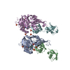
| ||||||||
|---|---|---|---|---|---|---|---|---|---|
| 1 | 
| ||||||||
| 2 | 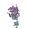
| ||||||||
| Unit cell |
|
-Components
| #1: Protein | Mass: 41744.570 Da / Num. of mol.: 2 / Fragment: CATALYTIC DOMAIN, RESIDUES 196-565 Source method: isolated from a genetically manipulated source Source: (gene. exp.) HOMO SAPIENS (human) / Production host:  References: UniProt: Q9UK80, EC: 3.1.2.15, ubiquitinyl hydrolase 1 #2: Protein | Mass: 17200.750 Da / Num. of mol.: 2 / Fragment: LINEAR DIUBIQUITIN ALDEHYDE, RESIDUES 1-152 / Mutation: YES Source method: isolated from a genetically manipulated source Details: NON-CLEAVABLE LINEAR DIUBIQUITIN ALDEHYDE WITH G76H MUTATION Source: (gene. exp.) HOMO SAPIENS (human) / Plasmid: PTYB2 / Production host: #3: Chemical |   Mass: 65.409 Da / Num. of mol.: 2 / Source method: obtained synthetically / Formula: Zn Mass: 65.409 Da / Num. of mol.: 2 / Source method: obtained synthetically / Formula: Zn#4: Chemical | ChemComp-SO4 /   Mass: 96.063 Da / Num. of mol.: 12 / Source method: obtained synthetically / Formula: SO4 Mass: 96.063 Da / Num. of mol.: 12 / Source method: obtained synthetically / Formula: SO4#5: Water | ChemComp-HOH / |  Mass: 18.015 Da / Num. of mol.: 26 / Source method: isolated from a natural source / Formula: H2O Mass: 18.015 Da / Num. of mol.: 26 / Source method: isolated from a natural source / Formula: H2OCompound details | ENGINEERED | Nonpolymer details | AMINO-ACETALDEHY | |
|---|
-Experimental details
-Experiment
| Experiment | Method: X-RAY DIFFRACTION / Number of used crystals: 1 |
|---|
- Sample preparation
| Crystal | Density Matthews: 2.36 Å3/Da / Density % sol: 48 % / Description: NONE |
|---|---|
| Crystal grow | pH: 7.4 / Details: 15% PEG8000, 0.2 M NH4SO4, pH 7.4 |
-Data collection
| Diffraction | Mean temperature: 100 K |
|---|---|
| Diffraction source | Source: SYNCHROTRON / Site: ESRF  / Beamline: ID23-2 / Wavelength: 1 / Beamline: ID23-2 / Wavelength: 1 |
| Detector | Type: MARRESEARCH / Detector: CCD / Date: May 9, 2009 |
| Radiation | Protocol: SINGLE WAVELENGTH / Monochromatic (M) / Laue (L): M / Scattering type: x-ray |
| Radiation wavelength | Wavelength: 1 Å / Relative weight: 1 |
| Reflection | Resolution: 2.7→49.8 Å / Num. obs: 24840 / % possible obs: 91.1 % / Observed criterion σ(I): 2 / Redundancy: 2.9 % / Biso Wilson estimate: 42.3 Å2 / Rmerge(I) obs: 0.12 / Net I/σ(I): 8.8 |
| Reflection shell | Resolution: 2.7→2.85 Å / Redundancy: 3 % / Rmerge(I) obs: 0.47 / Mean I/σ(I) obs: 2.4 / % possible all: 86.2 |
- Processing
| Software |
| ||||||||||||||||||||||||||||||||||||||||||||||||||||||||||||||||||||||||||||||||||||||||||||||||||||||||||||||||||||||||||||||||||||||||||||||||||||||||||||||||||||||||||||||||||||||
|---|---|---|---|---|---|---|---|---|---|---|---|---|---|---|---|---|---|---|---|---|---|---|---|---|---|---|---|---|---|---|---|---|---|---|---|---|---|---|---|---|---|---|---|---|---|---|---|---|---|---|---|---|---|---|---|---|---|---|---|---|---|---|---|---|---|---|---|---|---|---|---|---|---|---|---|---|---|---|---|---|---|---|---|---|---|---|---|---|---|---|---|---|---|---|---|---|---|---|---|---|---|---|---|---|---|---|---|---|---|---|---|---|---|---|---|---|---|---|---|---|---|---|---|---|---|---|---|---|---|---|---|---|---|---|---|---|---|---|---|---|---|---|---|---|---|---|---|---|---|---|---|---|---|---|---|---|---|---|---|---|---|---|---|---|---|---|---|---|---|---|---|---|---|---|---|---|---|---|---|---|---|---|---|
| Refinement | Method to determine structure: MOLECULAR REPLACEMENT Starting model: PDB ENTRIES 2HD5 AND 1UBQ Resolution: 2.7→45 Å / Cor.coef. Fo:Fc: 0.9 / Cor.coef. Fo:Fc free: 0.829 / SU B: 14.82 / SU ML: 0.306 / Cross valid method: THROUGHOUT / ESU R Free: 0.438 / Stereochemistry target values: MAXIMUM LIKELIHOOD Details: HYDROGENS HAVE BEEN ADDED IN THE RIDING POSITIONS. HYDROGENS HAVE BEEN USED IF PRESENT IN THE INPUT. U VALUES REFINED INDIVIDUALLY. COVALENT LINK BETWEEN C221 IN CHAIN A,E AND GLZ152 IN CHAIN B,F.
| ||||||||||||||||||||||||||||||||||||||||||||||||||||||||||||||||||||||||||||||||||||||||||||||||||||||||||||||||||||||||||||||||||||||||||||||||||||||||||||||||||||||||||||||||||||||
| Solvent computation | Ion probe radii: 0.8 Å / Shrinkage radii: 0.8 Å / VDW probe radii: 1.2 Å / Solvent model: MASK | ||||||||||||||||||||||||||||||||||||||||||||||||||||||||||||||||||||||||||||||||||||||||||||||||||||||||||||||||||||||||||||||||||||||||||||||||||||||||||||||||||||||||||||||||||||||
| Displacement parameters | Biso mean: 20.315 Å2
| ||||||||||||||||||||||||||||||||||||||||||||||||||||||||||||||||||||||||||||||||||||||||||||||||||||||||||||||||||||||||||||||||||||||||||||||||||||||||||||||||||||||||||||||||||||||
| Refinement step | Cycle: LAST / Resolution: 2.7→45 Å
| ||||||||||||||||||||||||||||||||||||||||||||||||||||||||||||||||||||||||||||||||||||||||||||||||||||||||||||||||||||||||||||||||||||||||||||||||||||||||||||||||||||||||||||||||||||||
| Refine LS restraints |
|