 Movie
Movie Controller

[English] 日本語
 Yorodumi
- PDB-4pn1: Structure of S. pombe Pct1 RNA triphosphatase in complex with the... -
+ Open data

- Basic information
| Entry | Database: PDB / ID: 4pn1 | ||||||
|---|---|---|---|---|---|---|---|
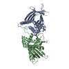
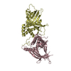
| Title | Structure of S. pombe Pct1 RNA triphosphatase in complex with the Spt5 CTD | ||||||
 Components Components |
| ||||||
 Keywords Keywords | HYDROLASE/TRANSCRIPTION REGULATOR / mRNA triphosphatase / hydrolase / polynucleotide 5' triphosphatase / mRNA processing / mRNA capping / dimer / transcription elongation factor / HYDROLASE-TRANSCRIPTION REGULATOR complex | ||||||
| Function / homology |  Function and homology information Function and homology informationpolynucleotide 5'-phosphatase activity / mRNA 5'-phosphatase / mRNA 5'-triphosphate monophosphatase activity / RNA polymerase II C-terminal domain binding / 7-methylguanosine mRNA capping / ATP hydrolysis activity / nucleus Similarity search - Function | ||||||
| Biological species |  | ||||||
| Method |  X-RAY DIFFRACTION / SYNCHROTRON / MOLECULAR REPLACEMENT / Resolution: 2.803 Å X-RAY DIFFRACTION / SYNCHROTRON / MOLECULAR REPLACEMENT / Resolution: 2.803 Å | ||||||
 Authors Authors | Lima, C.D. / Doamekpor, S.K. | ||||||
| Funding support |  United States, 1items United States, 1items
| ||||||
 Citation Citation | Journal: Rna / Year: 2015 Title: Fission yeast RNA triphosphatase reads an Spt5 CTD code. Authors: Doamekpor, S.K. / Schwer, B. / Sanchez, A.M. / Shuman, S. / Lima, C.D. | ||||||
| History |
|
- Structure visualization
| Structure viewer | Molecule: MolmilJmol/JSmol |
|---|
- Downloads & links
-Download
| PDBx/mmCIF format | 4pn1.cif.gz | 227.5 KB | Display | PDBx/mmCIF format |
|---|---|---|---|---|
| PDB format | pdb4pn1.ent.gz | 183.5 KB | Display | PDB format |
| PDBx/mmJSON format | 4pn1.json.gz | Tree view | PDBx/mmJSON format | |
| Others |  Other downloads Other downloads |
-Validation report
| Arichive directory | https://data.pdbj.org/pub/pdb/validation_reports/pn/4pn1ftp://data.pdbj.org/pub/pdb/validation_reports/pn/4pn1 | HTTPS FTP |
|---|
-Related structure data
| Related structure data |  4pn0SC S: Starting model for refinement C: citing same article ( |
|---|---|
| Similar structure data |


-Links
 PDBj
PDBj
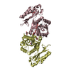
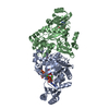
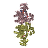
- Assembly
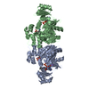
| Deposited unit | 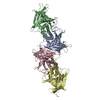
| ||||||||
|---|---|---|---|---|---|---|---|---|---|
| 1 | 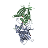
| ||||||||
| 2 | 
| ||||||||
| 3 |
| ||||||||
| Unit cell |
|
-Components
| #1: Protein | Mass: 35586.305 Da / Num. of mol.: 4 Source method: isolated from a genetically manipulated source Source: (gene. exp.) Gene: pct1 / Plasmid: pSMT3 / Details (production host): his-tagged SUMO fusion / Production host:  #2: Protein/peptide | Mass: 1906.022 Da / Num. of mol.: 4 / Source method: obtained synthetically Details: synthetic peptide derived from the repeating sequence found at the C-terminal domain of Spt5 #3: Chemical | ChemComp-FMT /   Mass: 46.025 Da / Num. of mol.: 6 / Source method: obtained synthetically / Formula: CH2O2 Mass: 46.025 Da / Num. of mol.: 6 / Source method: obtained synthetically / Formula: CH2O2#4: Chemical |   Mass: 62.068 Da / Num. of mol.: 2 / Source method: obtained synthetically / Formula: C2H6O2 Mass: 62.068 Da / Num. of mol.: 2 / Source method: obtained synthetically / Formula: C2H6O2#5: Water | ChemComp-HOH / |  Mass: 18.015 Da / Num. of mol.: 125 / Source method: isolated from a natural source / Formula: H2O Mass: 18.015 Da / Num. of mol.: 125 / Source method: isolated from a natural source / Formula: H2O |
|---|
-Experimental details
-Experiment
| Experiment | Method: X-RAY DIFFRACTION / Number of used crystals: 1 |
|---|
- Sample preparation
| Crystal | Density Matthews: 3.02 Å3/Da / Density % sol: 59.31 % |
|---|---|
| Crystal grow | Temperature: 291 K / Method: vapor diffusion, hanging drop Details: 3.0 M sodium formate cryo-protected in 3.2 M sodium formate and 20% (v/v) ethylene glycol |
-Data collection
| Diffraction | Mean temperature: 100 K |
|---|---|
| Diffraction source | Source: SYNCHROTRON / Site: APS / Beamline: 24-ID-E / Wavelength: 0.979 Å |
| Detector | Type: ADSC QUANTUM 315 / Detector: CCD / Date: Feb 1, 2010 |
| Radiation | Protocol: SINGLE WAVELENGTH / Monochromatic (M) / Laue (L): M / Scattering type: x-ray |
| Radiation wavelength | Wavelength: 0.979 Å / Relative weight: 1 |
| Reflection | Resolution: 2.8→50 Å / Num. obs: 49568 / % possible obs: 99.1 % / Redundancy: 4.9 % / Biso Wilson estimate: 46.1 Å2 / Rmerge(I) obs: 0.113 / Net I/σ(I): 13.8 |
| Reflection shell | Resolution: 2.8→2.9 Å / Redundancy: 4 % / Rmerge(I) obs: 0.43 / Mean I/σ(I) obs: 2.6 / % possible all: 97.7 |
- Processing
| Software | Name: PHENIX / Version: (phenix.refine: 1.8.2_1309) / Classification: refinement | |||||||||||||||||||||||||||||||||||||||||||||||||||||||||||||||||||||||||||||||||||||||||||||||||||||||||||||||||||||||
|---|---|---|---|---|---|---|---|---|---|---|---|---|---|---|---|---|---|---|---|---|---|---|---|---|---|---|---|---|---|---|---|---|---|---|---|---|---|---|---|---|---|---|---|---|---|---|---|---|---|---|---|---|---|---|---|---|---|---|---|---|---|---|---|---|---|---|---|---|---|---|---|---|---|---|---|---|---|---|---|---|---|---|---|---|---|---|---|---|---|---|---|---|---|---|---|---|---|---|---|---|---|---|---|---|---|---|---|---|---|---|---|---|---|---|---|---|---|---|---|---|
| Refinement | Method to determine structure: MOLECULAR REPLACEMENT Starting model: 4PN0 Resolution: 2.803→48.362 Å / SU ML: 0.38 / Cross valid method: FREE R-VALUE / σ(F): 1.43 / Phase error: 23.79 / Stereochemistry target values: ML
| |||||||||||||||||||||||||||||||||||||||||||||||||||||||||||||||||||||||||||||||||||||||||||||||||||||||||||||||||||||||
| Solvent computation | Shrinkage radii: 0.9 Å / VDW probe radii: 1.11 Å / Solvent model: FLAT BULK SOLVENT MODEL | |||||||||||||||||||||||||||||||||||||||||||||||||||||||||||||||||||||||||||||||||||||||||||||||||||||||||||||||||||||||
| Displacement parameters | Biso mean: 56.2 Å2 | |||||||||||||||||||||||||||||||||||||||||||||||||||||||||||||||||||||||||||||||||||||||||||||||||||||||||||||||||||||||
| Refinement step | Cycle: LAST / Resolution: 2.803→48.362 Å
| |||||||||||||||||||||||||||||||||||||||||||||||||||||||||||||||||||||||||||||||||||||||||||||||||||||||||||||||||||||||
| Refine LS restraints |
| |||||||||||||||||||||||||||||||||||||||||||||||||||||||||||||||||||||||||||||||||||||||||||||||||||||||||||||||||||||||
| LS refinement shell |
|
