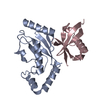
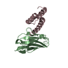
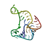
Rad6-Rad18 complex / positive regulation of chromosome segregation / Y-form DNA binding / nuclear inclusion body / DNA damage tolerance / polyubiquitin modification-dependent protein binding / protein monoubiquitination / protein autoubiquitination / replication fork / Recognition of DNA damage by PCNA-containing replication complex ...Rad6-Rad18 complex / positive regulation of chromosome segregation / Y-form DNA binding / nuclear inclusion body / DNA damage tolerance / polyubiquitin modification-dependent protein binding / protein monoubiquitination / protein autoubiquitination / replication fork / Recognition of DNA damage by PCNA-containing replication complex / RING-type E3 ubiquitin transferase / ubiquitin protein ligase activity / single-stranded DNA binding / E3 ubiquitin ligases ubiquitinate target proteins / site of double-strand break / damaged DNA binding / nuclear body / DNA repair / DNA damage response / ubiquitin protein ligase binding / centrosome / protein-containing complex binding / zinc ion binding / nucleoplasm / identical protein binding / nucleus / cytoplasm Similarity search - Function
E3 ubiquitin-protein ligase Rad18 / Rad18-like CCHC zinc finger / Zinc finger, C3HC4 type (RING finger) / SAP domain superfamily / SAP motif profile. / SAP domain / Putative DNA-binding (bihelical) motif predicted to be involved in chromosomal organisation / SAP domain / Rad18, zinc finger UBZ4-type / Zinc finger UBZ4-type profile. ...E3 ubiquitin-protein ligase Rad18 / Rad18-like CCHC zinc finger / Zinc finger, C3HC4 type (RING finger) / SAP domain superfamily / SAP motif profile. / SAP domain / Putative DNA-binding (bihelical) motif predicted to be involved in chromosomal organisation / SAP domain / Rad18, zinc finger UBZ4-type / Zinc finger UBZ4-type profile. / Zinc/RING finger domain, C3HC4 (zinc finger) / Herpes Virus-1 / Zinc finger, RING-type, conserved site / Zinc finger RING-type signature. / Ring finger / Zinc finger RING-type profile. / Zinc finger, RING-type / Zinc finger, RING/FYVE/PHD-type / 2-Layer Sandwich / Alpha Beta Similarity search - Domain/homology
Protocol: SINGLE WAVELENGTH / Monochromatic (M) / Laue (L): M / Scattering type: x-ray
Radiation wavelength
Wavelength: 1.2828 Å / Relative weight: 1
Reflection
Resolution: 1.8→42.84 Å / Num. obs: 16560 / % possible obs: 99.9 % / Observed criterion σ(I): 3 / Redundancy: 17.3 % / Biso Wilson estimate: 18.4 Å2 / Rmerge(I) obs: 0.09 / Net I/σ(I): 24.3
Reflection shell
Resolution: 1.8→1.89 Å / Redundancy: 5.8 % / Rmerge(I) obs: 0.62 / Mean I/σ(I) obs: 2.5 / % possible all: 99.2
-
Processing
Software
Name
Version
Classification
XDS
datareduction
SCALA
datascaling
SHELX
phasing
PHASER
phasing
REFMAC
5.5.0099
refinement
Refinement
Method to determine structure: SAD Starting model: NONE Resolution: 1.8→42.86 Å / Cor.coef. Fo:Fc: 0.957 / Cor.coef. Fo:Fc free: 0.932 / SU B: 5.874 / SU ML: 0.082 / Cross valid method: THROUGHOUT / ESU R: 0.127 / ESU R Free: 0.127 / Stereochemistry target values: MAXIMUM LIKELIHOOD / Details: HYDROGENS HAVE BEEN ADDED IN THE RIDING POSITIONS.
Rfactor
Num. reflection
% reflection
Selection details
Rfree
0.22311
828
5 %
RANDOM
Rwork
0.17505
-
-
-
obs
0.1774
15732
99.92 %
-
Solvent computation
Ion probe radii: 0.8 Å / Shrinkage radii: 0.8 Å / VDW probe radii: 1.4 Å / Solvent model: MASK
 Movie
Movie Controller


 Open data
 Basic information
 Components
 Keywords
 Function and homology information
 HOMO SAPIENS (human)
 X-RAY DIFFRACTION /
  Authors
 Citation
 Structure visualization
 Downloads & links
 Other downloads


 PDBj
PDBj


 Assembly
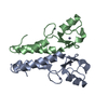


 Mass: 65.409 Da / Num. of mol.: 4 / Source method: obtained synthetically / Formula: Zn
 Mass: 18.015 Da / Num. of mol.: 102 / Source method: isolated from a natural source / Formula: H2O
 Sample preparation
 / Beamline: ID23-1 / Wavelength: 1.2828
  Processing