 Movie
Movie Controller

[English] 日本語
 Yorodumi
- PDB-2x7p: The Conserved Candida albicans CA3427 Gene Product Defines a New ... -
+ Open data

- Basic information
| Entry | Database: PDB / ID: 2x7p | ||||||
|---|---|---|---|---|---|---|---|
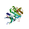
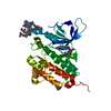
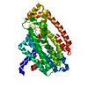
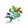
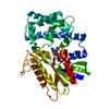
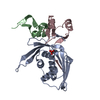
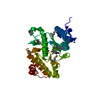
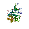
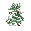
| Title | The Conserved Candida albicans CA3427 Gene Product Defines a New Family of Proteins Exhibiting the Generic Periplasmic Binding Protein Structural Fold | ||||||
 Components Components | POSSIBLE THIAMINE BIOSYNTHESIS ENZYME | ||||||
 Keywords Keywords | UNKNOWN FUNCTION | ||||||
| Function / homology |  Function and homology information Function and homology information | ||||||
| Biological species |   CANDIDA ALBICANS (yeast) CANDIDA ALBICANS (yeast) | ||||||
| Method | X-RAY DIFFRACTION / SYNCHROTRON / MAD / Resolution: 2.34 Å | ||||||
 Authors Authors | Santini, S. / Monchois, V. / Mouz, N. / Rousselle, T. / Claverie, J.M. / Abergel, C. | ||||||
 Citation Citation | Journal: Plos One / Year: 2011 Title: The Conserved Candida Albicans Ca3427 Gene Product Defines a New Family of Proteins Exhibiting the Generic Periplasmic Binding Protein Structural Fold Authors: Santini, S. / Claverie, J.M. / Mouz, N. / Rousselle, T. / Maza, C. / Monchois, V. / Abergel, C. | ||||||
| History |
|
- Structure visualization
| Structure viewer | Molecule: MolmilJmol/JSmol |
|---|
- Downloads & links
-Download
| PDBx/mmCIF format | 2x7p.cif.gz | 76 KB | Display | PDBx/mmCIF format |
|---|---|---|---|---|
| PDB format | pdb2x7p.ent.gz | 59.7 KB | Display | PDB format |
| PDBx/mmJSON format | 2x7p.json.gz | Tree view | PDBx/mmJSON format | |
| Others |  Other downloads Other downloads |
-Validation report
| Arichive directory | https://data.pdbj.org/pub/pdb/validation_reports/x7/2x7pftp://data.pdbj.org/pub/pdb/validation_reports/x7/2x7p | HTTPS FTP |
|---|
-Related structure data


-Links
 PDBj
PDBj


- Assembly
| Deposited unit | 
| ||||||||
|---|---|---|---|---|---|---|---|---|---|
| 1 |
| ||||||||
| Unit cell |
|
-Components
-Protein , 1 types, 1 molecules A
| #1: Protein | Mass: 36371.113 Da / Num. of mol.: 1 / Mutation: YES Source method: isolated from a genetically manipulated source Source: (gene. exp.) CANDIDA ALBICANS (yeast) / Strain: NIH3147 / Plasmid: PSF04, PQE80 / Production host:  ESCHERICHIA COLI (E. coli) / Strain (production host): K-12 / References: UniProt: Q59X88 ESCHERICHIA COLI (E. coli) / Strain (production host): K-12 / References: UniProt: Q59X88 |
|---|
-Non-polymers , 7 types, 151 molecules 


| #2: Chemical | ChemComp-CA /  Mass: 40.078 Da / Num. of mol.: 1 / Source method: obtained synthetically / Formula: Ca Mass: 40.078 Da / Num. of mol.: 1 / Source method: obtained synthetically / Formula: Ca | ||||||||
|---|---|---|---|---|---|---|---|---|---|
| #3: Chemical | ChemComp-CL / Chloride Mass: 35.453 Da / Num. of mol.: 1 / Source method: obtained synthetically / Formula: Cl Mass: 35.453 Da / Num. of mol.: 1 / Source method: obtained synthetically / Formula: Cl | ||||||||
| #4: Chemical | Glycerol Mass: 92.094 Da / Num. of mol.: 2 / Source method: obtained synthetically / Formula: C3H8O3 Mass: 92.094 Da / Num. of mol.: 2 / Source method: obtained synthetically / Formula: C3H8O3#5: Chemical | ChemComp-AE3 / | 2-(2-Ethoxyethoxy)ethanol Mass: 134.174 Da / Num. of mol.: 1 / Source method: obtained synthetically / Formula: C6H14O3 Mass: 134.174 Da / Num. of mol.: 1 / Source method: obtained synthetically / Formula: C6H14O3#6: Chemical | ChemComp-ACY / Acetic acid Mass: 60.052 Da / Num. of mol.: 5 / Source method: obtained synthetically / Formula: C2H4O2 Mass: 60.052 Da / Num. of mol.: 5 / Source method: obtained synthetically / Formula: C2H4O2#7: Chemical | ChemComp-CO2 / | Carbon dioxide Mass: 44.010 Da / Num. of mol.: 1 / Source method: obtained synthetically / Formula: CO2 Mass: 44.010 Da / Num. of mol.: 1 / Source method: obtained synthetically / Formula: CO2#8: Water | ChemComp-HOH / | WaterMass: 18.015 Da / Num. of mol.: 140 / Source method: isolated from a natural source / Formula: H2O |
-Experimental details
-Experiment
| Experiment | Method: X-RAY DIFFRACTION / Number of used crystals: 1 |
|---|
- Sample preparation
| Crystal | Density Matthews: 2.44 Å3/Da / Density % sol: 49.52 % / Description: NONE |
|---|---|
| Crystal grow | Details: 21 % PEG8000, CALCIUM ACETATE 0.2 M, TRIS 0.1 M, 30 % GLYCEROL PH 7.0 |
-Data collection
| Diffraction | Mean temperature: 100 K | |||||||||
|---|---|---|---|---|---|---|---|---|---|---|
| Diffraction source | Source: SYNCHROTRON / Site: ESRF  / Beamline: BM30A / Wavelength: 0.979774, 0.979958 / Beamline: BM30A / Wavelength: 0.979774, 0.979958 | |||||||||
| Detector | Type: MARRESEARCH / Date: Jul 3, 2004 | |||||||||
| Radiation | Protocol: MAD / Monochromatic (M) / Laue (L): M / Scattering type: x-ray | |||||||||
| Radiation wavelength |
| |||||||||
| Reflection | Resolution: 2.34→23.88 Å / Num. obs: 13442 / % possible obs: 99.1 % / Observed criterion σ(I): 10 / Redundancy: 5.5 % / Biso Wilson estimate: 24.14 Å2 / Rmerge(I) obs: 0.08 / Net I/σ(I): 4.1 | |||||||||
| Reflection shell | Resolution: 2.34→2.47 Å / Redundancy: 3 % / Rmerge(I) obs: 0.31 / Mean I/σ(I) obs: 3 / % possible all: 99.1 |
- Processing
| Software |
| ||||||||||||||||||||||||||||||||||||||||||
|---|---|---|---|---|---|---|---|---|---|---|---|---|---|---|---|---|---|---|---|---|---|---|---|---|---|---|---|---|---|---|---|---|---|---|---|---|---|---|---|---|---|---|---|
| Refinement | Method to determine structure: MAD Starting model: NONE Resolution: 2.34→23.88 Å / SU ML: 0.28 / σ(F): 2.46 / Phase error: 21.89 / Stereochemistry target values: ML Details: DISORDERED SIDE CHAIN ARE MODELED WITH A OCCUPANCY LESS THAN 1 WITHOUT ALTERNATE CONFORMATION.
| ||||||||||||||||||||||||||||||||||||||||||
| Solvent computation | Shrinkage radii: 0.9 Å / VDW probe radii: 1.11 Å / Solvent model: FLAT BULK SOLVENT MODEL / Bsol: 40.03 Å2 / ksol: 0.4 e/Å3 | ||||||||||||||||||||||||||||||||||||||||||
| Displacement parameters | Biso mean: 22.75 Å2
| ||||||||||||||||||||||||||||||||||||||||||
| Refinement step | Cycle: LAST / Resolution: 2.34→23.88 Å
| ||||||||||||||||||||||||||||||||||||||||||
| Refine LS restraints |
| ||||||||||||||||||||||||||||||||||||||||||
| LS refinement shell |
|
