- PDB-2wbn: Crystal structure of the g2p (large terminase) nuclease domain fr... -
+
Open data
ID or keywords:
Loading...
-
Basic information
Entry
Database: PDB / ID: 2wbn
Title
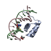
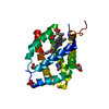
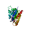
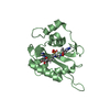
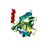
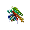
Crystal structure of the g2p (large terminase) nuclease domain from the bacteriophage SPP1
Components
TERMINASE LARGE SUBUNIT
Keywords
VIRAL PROTEIN / LARGE TERMINASE / NUCLEASE / DNA PACKAGING
Function / homology
Function and homology information
viral terminase, large subunit / viral DNA genome packaging / nuclease activity / chromosome organization / Hydrolases; Acting on acid anhydrides; Acting on acid anhydrides to facilitate cellular and subcellular movement / endonuclease activity / Hydrolases; Acting on ester bonds / ATP hydrolysis activity / ATP binding / metal ion binding Similarity search - Function
Phage terminase large subunit, C-terminal / Terminase, large subunit SPP1-like / Nucleotidyltransferase; domain 5 - #280 / Terminase RNAseH like domain / Phage terminase large subunit, N-terminal / : / Phage terminase large subunit / Bacteriophage terminase, large subunit / Nucleotidyltransferase; domain 5 / P-loop containing nucleoside triphosphate hydrolase ...Phage terminase large subunit, C-terminal / Terminase, large subunit SPP1-like / Nucleotidyltransferase; domain 5 - #280 / Terminase RNAseH like domain / Phage terminase large subunit, N-terminal / : / Phage terminase large subunit / Bacteriophage terminase, large subunit / Nucleotidyltransferase; domain 5 / P-loop containing nucleoside triphosphate hydrolase / 2-Layer Sandwich / Alpha Beta Similarity search - Domain/homology
 Movie
Movie Controller

 Yorodumi
 Open data
 Basic information
 Components
 Keywords
 Function and homology information
 BACILLUS PHAGE SPP1 (virus)
 X-RAY DIFFRACTION /
  Authors
 Citation
 Structure visualization
 Downloads & links
 Other downloads
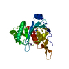


 PDBj
PDBj Assembly

 Mass: 18.015 Da / Num. of mol.: 127 / Source method: isolated from a natural source / Formula: H2O
 Sample preparation
 / Beamline: BM14 / Wavelength: 0.9184
  Processing