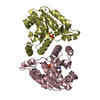
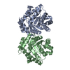
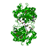
The biological assembly is a homo-dimer generated by a diagonal two-fold rotation axis of the space group [x,y,z; y,x,-z]. The homo-dimer has a very high complexation significance score (CSS) of 0.967 [PISA web site results at EBI]. The other four components of the unit cell are generated by operation of the three-fold screw axis 3(1).
-
Components
#1: Protein
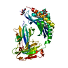
UncharacterizedproteinygiD
Mass: 30492.955 Da / Num. of mol.: 1 Source method: isolated from a genetically manipulated source Source: (gene. exp.) Escherichia coli (E. coli) / Strain: K12 / Gene: ygiD, b3039, JW3007 / Plasmid: PCR2.1-TOPO / Production host: Escherichia coli (E. coli) / References: UniProt: P24197
Type: MARMOSAIC 300 mm CCD / Detector: CCD / Date: Aug 6, 2006 / Details: ROSENBAUM
Radiation
Monochromator: SI CHANNEL 220 / Protocol: SINGLE WAVELENGTH / Monochromatic (M) / Laue (L): M / Scattering type: x-ray
Radiation wavelength
Wavelength: 0.97243 Å / Relative weight: 1
Reflection
Resolution: 2.27→72.74 Å / Num. obs: 16342 / % possible obs: 99.6 % / Observed criterion σ(I): 1 / Redundancy: 20.7 % / Rsym value: 0.096 / Net I/σ(I): 18.1
Reflection shell
Resolution: 2.27→2.35 Å / Redundancy: 20.7 % / Mean I/σ(I) obs: 5.63 / Rsym value: 0.63 / % possible all: 100
-
Processing
Software
Name
Version
Classification
SCA2STRUCTURE
modelbuilding
REFMAC
5.2.0019
refinement
SERGUI
datacollection
HKL-2000
datareduction
SCALEPACK
datascaling
SCA2STRUCTURE
phasing
Refinement
Method to determine structure: SAD / Resolution: 2.27→72.74 Å / Cor.coef. Fo:Fc: 0.936 / Cor.coef. Fo:Fc free: 0.918 / SU B: 6.833 / SU ML: 0.169 / Cross valid method: THROUGHOUT / ESU R: 0.27 / ESU R Free: 0.218 / Stereochemistry target values: MAXIMUM LIKELIHOOD / Details: HYDROGENS HAVE BEEN ADDED IN THE RIDING POSITIONS
Rfactor
Num. reflection
% reflection
Selection details
Rfree
0.2667
825
5.1 %
RANDOM
Rwork
0.23457
-
-
-
obs
0.23616
15481
99.4 %
-
Solvent computation
Ion probe radii: 0.8 Å / Shrinkage radii: 0.8 Å / VDW probe radii: 1.2 Å / Solvent model: MASK
Displacement parameters
Biso mean: 48.677 Å2
Baniso -1
Baniso -2
Baniso -3
1-
-0.98 Å2
-0.49 Å2
0 Å2
2-
-
-0.98 Å2
0 Å2
3-
-
-
1.47 Å2
Refinement step
Cycle: LAST / Resolution: 2.27→72.74 Å
Protein
Nucleic acid
Ligand
Solvent
Total
Num. atoms
1869
0
3
51
1923
Refine LS restraints
Refine-ID
Type
Dev ideal
Dev ideal target
Number
X-RAY DIFFRACTION
r_bond_refined_d
0.016
0.022
1928
X-RAY DIFFRACTION
r_angle_refined_deg
1.703
1.95
2638
X-RAY DIFFRACTION
r_dihedral_angle_1_deg
7.772
5
241
X-RAY DIFFRACTION
r_dihedral_angle_2_deg
38.492
23.896
77
X-RAY DIFFRACTION
r_dihedral_angle_3_deg
15.814
15
281
X-RAY DIFFRACTION
r_dihedral_angle_4_deg
22.464
15
7
X-RAY DIFFRACTION
r_chiral_restr
0.122
0.2
286
X-RAY DIFFRACTION
r_gen_planes_refined
0.006
0.02
1484
X-RAY DIFFRACTION
r_nbd_refined
0.213
0.2
895
X-RAY DIFFRACTION
r_nbtor_refined
0.308
0.2
1264
X-RAY DIFFRACTION
r_xyhbond_nbd_refined
0.142
0.2
69
X-RAY DIFFRACTION
r_metal_ion_refined
0.055
0.2
2
X-RAY DIFFRACTION
r_symmetry_vdw_refined
0.228
0.2
29
X-RAY DIFFRACTION
r_symmetry_hbond_refined
0.245
0.2
4
X-RAY DIFFRACTION
r_symmetry_metal_ion_refined
0.482
0.2
1
X-RAY DIFFRACTION
r_mcbond_it
1.022
1.5
1236
X-RAY DIFFRACTION
r_mcangle_it
1.554
2
1942
X-RAY DIFFRACTION
r_scbond_it
2.197
3
815
X-RAY DIFFRACTION
r_scangle_it
3.008
4.5
696
LS refinement shell
Resolution: 2.27→2.33 Å / Total num. of bins used: 20
Rfactor
Num. reflection
% reflection
Rfree
0.355
61
-
Rwork
0.348
1101
-
obs
-
-
97.24 %
+
About Yorodumi
-
News
-
Feb 9, 2022. New format data for meta-information of EMDB entries
New format data for meta-information of EMDB entries
Version 3 of the EMDB header file is now the official format.
The previous official version 1.9 will be removed from the archive.
In the structure databanks used in Yorodumi, some data are registered as the other names, "COVID-19 virus" and "2019-nCoV". Here are the details of the virus and the list of structure data.
Jan 31, 2019. EMDB accession codes are about to change! (news from PDBe EMDB page)
EMDB accession codes are about to change! (news from PDBe EMDB page)
The allocation of 4 digits for EMDB accession codes will soon come to an end. Whilst these codes will remain in use, new EMDB accession codes will include an additional digit and will expand incrementally as the available range of codes is exhausted. The current 4-digit format prefixed with “EMD-” (i.e. EMD-XXXX) will advance to a 5-digit format (i.e. EMD-XXXXX), and so on. It is currently estimated that the 4-digit codes will be depleted around Spring 2019, at which point the 5-digit format will come into force.
The EM Navigator/Yorodumi systems omit the EMD- prefix.
Related info.:Q: What is EMD? / ID/Accession-code notation in Yorodumi/EM Navigator
Yorodumi is a browser for structure data from EMDB, PDB, SASBDB, etc.
This page is also the successor to EM Navigator detail page, and also detail information page/front-end page for Omokage search.
The word "yorodu" (or yorozu) is an old Japanese word meaning "ten thousand". "mi" (miru) is to see.
Related info.:EMDB / PDB / SASBDB / Comparison of 3 databanks / Yorodumi Search / Aug 31, 2016. New EM Navigator & Yorodumi / Yorodumi Papers / Jmol/JSmol / Function and homology information / Changes in new EM Navigator and Yorodumi
 Movie
Movie Controller

 Yorodumi
 Open data
 Basic information
 Components
 Keywords
 Function and homology information
 X-RAY DIFFRACTION /
  Authors
 Citation
 Structure visualization
 Downloads & links
 Other downloads
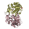


 PDBj
PDBj Assembly
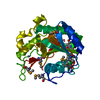

 Mass: 65.409 Da / Num. of mol.: 3 / Source method: obtained synthetically / Formula: Zn
 Mass: 18.015 Da / Num. of mol.: 51 / Source method: isolated from a natural source / Formula: H2O
 Sample preparation
 / Beamline: 22-ID / Wavelength: 0.97243 Å
 Processing