 Movie
Movie Controller

[English] 日本語
 Yorodumi
- PDB-2pk8: Crystal structure of an uncharacterized protein PF0899 from Pyroc... -
+ Open data

- Basic information
| Entry | Database: PDB / ID: 2pk8 | |||||||||
|---|---|---|---|---|---|---|---|---|---|---|
| Title | Crystal structure of an uncharacterized protein PF0899 from Pyrococcus furiosus | |||||||||
 Components Components | Uncharacterized protein PF0899 | |||||||||
 Keywords Keywords |  STRUCTURAL GENOMICS / UNKNOWN FUNCTION / PF0899 / PYROCOCCUS FURIOSUS / UNCHARACTERIZED PROTEIN / PSI / PROTEIN STRUCTURE INITIATIVE / SOUTHEAST COLLABORATORY FOR STRUCTURAL GENOMICS / SECSG STRUCTURAL GENOMICS / UNKNOWN FUNCTION / PF0899 / PYROCOCCUS FURIOSUS / UNCHARACTERIZED PROTEIN / PSI / PROTEIN STRUCTURE INITIATIVE / SOUTHEAST COLLABORATORY FOR STRUCTURAL GENOMICS / SECSG | |||||||||
| Function / homology | Putative type 4B encapsulin shell protein / Putative type 4B encapsulin shell protein superfamily / Putative type 4B encapsulin shell protein / hypothetical protein PF0899 domain / hypothetical protein PF0899 fold / 2-Layer Sandwich / Alpha Beta / GOLD (I) CYANIDE ION / Uncharacterized protein Function and homology information Function and homology information | |||||||||
| Biological species |   Pyrococcus furiosus DSM 3638 (archaea) Pyrococcus furiosus DSM 3638 (archaea) | |||||||||
| Method | X-RAY DIFFRACTION / SYNCHROTRON / MIRAS / Resolution: 1.85 Å | |||||||||
 Authors Authors | Liu, Z.J. / Tempel, W. / Chen, L. / Shah, A. / Lee, D. / Clancy-Kelley, L.L. / Dillard, B.D. / Rose, J.P. / Sugar, F.J. / Jenny Jr., F.E. ...Liu, Z.J. / Tempel, W. / Chen, L. / Shah, A. / Lee, D. / Clancy-Kelley, L.L. / Dillard, B.D. / Rose, J.P. / Sugar, F.J. / Jenny Jr., F.E. / Lee, H.S. / Izumi, M. / Shah, C. / Poole III, F.L. / Adams, M.W.W. / Richardson, J.S. / Richardson, D.C. / Wang, B.-C. / Southeast Collaboratory for Structural Genomics (SECSG) | |||||||||
 Citation Citation | Journal: Acta Crystallogr.,Sect.F / Year: 2007 Title: Structure of the hypothetical protein PF0899 from Pyrococcus furiosus at 1.85 A resolution. Authors: Kelley, L.L. / Dillard, B.D. / Tempel, W. / Chen, L. / Shaw, N. / Lee, D. / Newton, M.G. / Sugar, F.J. / Jenney, F.E. / Lee, H.S. / Shah, C. / Poole, F.L. / Adams, M.W. / Richardson, J.S. / ...Authors: Kelley, L.L. / Dillard, B.D. / Tempel, W. / Chen, L. / Shaw, N. / Lee, D. / Newton, M.G. / Sugar, F.J. / Jenney, F.E. / Lee, H.S. / Shah, C. / Poole, F.L. / Adams, M.W. / Richardson, J.S. / Richardson, D.C. / Liu, Z.J. / Wang, B.C. / Rose, J. | |||||||||
| History |
|
- Structure visualization
| Structure viewer | Molecule: MolmilJmol/JSmol |
|---|
- Downloads & links
-Download
| PDBx/mmCIF format | 2pk8.cif.gz | 33.2 KB | Display | PDBx/mmCIF format |
|---|---|---|---|---|
| PDB format | pdb2pk8.ent.gz | 22.1 KB | Display | PDB format |
| PDBx/mmJSON format | 2pk8.json.gz | Tree view | PDBx/mmJSON format | |
| Others |  Other downloads Other downloads |
-Validation report
| Arichive directory | https://data.pdbj.org/pub/pdb/validation_reports/pk/2pk8ftp://data.pdbj.org/pub/pdb/validation_reports/pk/2pk8 | HTTPS FTP |
|---|
-Related structure data
| Similar structure data | |
|---|---|
| Other databases |


-Links
 PDBj
PDBj- Assembly
| Deposited unit | 
| ||||||||
|---|---|---|---|---|---|---|---|---|---|
| 1 | 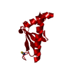
| ||||||||
| Unit cell |
| ||||||||
| Components on special symmetry positions |
|
-Components
| #1: Protein | Mass: 11570.343 Da / Num. of mol.: 1 Source method: isolated from a genetically manipulated source Details: THE PROTEIN WAS CLONED, EXPRESSED AND PURIFIED BY THE SECSG PYROCOCCUS PROTEIN PRODUCTION GROUP (M.W.W.ADAMS, P.S.BRERETON, M.IZUMI, F.E.JENNEY JR., H-S.LEE, F.L.POOLE II, C.SHAH, F.SUGAR) ...Details: THE PROTEIN WAS CLONED, EXPRESSED AND PURIFIED BY THE SECSG PYROCOCCUS PROTEIN PRODUCTION GROUP (M.W.W.ADAMS, P.S.BRERETON, M.IZUMI, F.E.JENNEY JR., H-S.LEE, F.L.POOLE II, C.SHAH, F.SUGAR) UNDER THE DIRECTION OF M.W.W.ADAMS. Source: (gene. exp.) Pyrococcus furiosus DSM 3638 (archaea) / Species: Pyrococcus furiosus / Strain: DSM 3638, JCM 8422, Vc1 / Gene: PF0899 / Plasmid: pET24D BAM / Production host:  Escherichia coli (E. coli) / Strain (production host): BL21 STAR DE3 PRIL / References: UniProt: Q8U2E0 Escherichia coli (E. coli) / Strain (production host): BL21 STAR DE3 PRIL / References: UniProt: Q8U2E0 |
|---|---|
| #2: Chemical | ChemComp-AUC /   Mass: 249.001 Da / Num. of mol.: 1 / Source method: obtained synthetically / Formula: C2AuN2 Mass: 249.001 Da / Num. of mol.: 1 / Source method: obtained synthetically / Formula: C2AuN2 |
| #3: Water | ChemComp-HOH / Water Mass: 18.015 Da / Num. of mol.: 38 / Source method: isolated from a natural source / Formula: H2O Mass: 18.015 Da / Num. of mol.: 38 / Source method: isolated from a natural source / Formula: H2O |
-Experimental details
-Experiment
| Experiment | Method: X-RAY DIFFRACTION / Number of used crystals: 1 |
|---|
- Sample preparation
| Crystal | Density Matthews: 2.32 Å3/Da / Density % sol: 47 % |
|---|---|
| Crystal grow | Temperature: 291 K / Method: vapor diffusion, sitting drop / pH: 3.9 Details: MODIFIED MICROBATCH USING 1 MICROLITER DROPS CONTAINING EQUAL VOLUMES OF PROTEIN CONCENTRATE (10 mg/mL) AND A PRECIPITANT SOLUTION CONTAINING 8% PEG 4000 IN 100mM SODIUM ACETATE, pH 3.9, ...Details: MODIFIED MICROBATCH USING 1 MICROLITER DROPS CONTAINING EQUAL VOLUMES OF PROTEIN CONCENTRATE (10 mg/mL) AND A PRECIPITANT SOLUTION CONTAINING 8% PEG 4000 IN 100mM SODIUM ACETATE, pH 3.9, TEMPERATURE 291K, VAPOR DIFFUSION, SITTING DROP |
-Data collection
| Diffraction | Mean temperature: 100 K |
|---|---|
| Diffraction source | Source: SYNCHROTRON / Site: APS  / Beamline: 22-ID / Wavelength: 1 Å / Beamline: 22-ID / Wavelength: 1 Å |
| Detector | Type: MARMOSAIC 225 mm CCD / Detector: CCD / Date: Nov 16, 2003 / Details: Rosenbaum |
| Radiation | Monochromator: SI CHANNEL 220 / Protocol: SINGLE WAVELENGTH / Monochromatic (M) / Laue (L): M / Scattering type: x-ray |
| Radiation wavelength | Wavelength: 1 Å / Relative weight: 1 |
| Reflection | Resolution: 1.85→20 Å / Num. obs: 9476 / % possible obs: 98.5 % / Observed criterion σ(I): -3 / Rsym value: 0.044 |
| Reflection shell | Resolution: 1.85→1.92 Å / Rsym value: 0.251 / % possible all: 92.4 |
- Processing
| Software |
| |||||||||||||||||||||||||||||||||||||||||||||||||||||||||||||||||||||||||||||||||||||||||||||||||||||||||||||||||||||||||||||||||||||||||||||||||||||||||||||||||||||||||||||||
|---|---|---|---|---|---|---|---|---|---|---|---|---|---|---|---|---|---|---|---|---|---|---|---|---|---|---|---|---|---|---|---|---|---|---|---|---|---|---|---|---|---|---|---|---|---|---|---|---|---|---|---|---|---|---|---|---|---|---|---|---|---|---|---|---|---|---|---|---|---|---|---|---|---|---|---|---|---|---|---|---|---|---|---|---|---|---|---|---|---|---|---|---|---|---|---|---|---|---|---|---|---|---|---|---|---|---|---|---|---|---|---|---|---|---|---|---|---|---|---|---|---|---|---|---|---|---|---|---|---|---|---|---|---|---|---|---|---|---|---|---|---|---|---|---|---|---|---|---|---|---|---|---|---|---|---|---|---|---|---|---|---|---|---|---|---|---|---|---|---|---|---|---|---|---|---|---|
| Refinement | Method to determine structure: MIRAS / Resolution: 1.85→19.81 Å / Cor.coef. Fo:Fc: 0.947 / Cor.coef. Fo:Fc free: 0.93 / SU B: 8.053 / SU ML: 0.116 / TLS residual ADP flag: LIKELY RESIDUAL / Cross valid method: THROUGHOUT / σ(F): 0 / σ(I): 0.257 / ESU R: 0.145 / ESU R Free: 0.137 / Stereochemistry target values: MAXIMUM LIKELIHOOD / Details: HYDROGENS HAVE BEEN ADDED IN THE RIDING POSITIONS
| |||||||||||||||||||||||||||||||||||||||||||||||||||||||||||||||||||||||||||||||||||||||||||||||||||||||||||||||||||||||||||||||||||||||||||||||||||||||||||||||||||||||||||||||
| Solvent computation | Ion probe radii: 0.8 Å / Shrinkage radii: 0.8 Å / VDW probe radii: 1.4 Å / Solvent model: MASK | |||||||||||||||||||||||||||||||||||||||||||||||||||||||||||||||||||||||||||||||||||||||||||||||||||||||||||||||||||||||||||||||||||||||||||||||||||||||||||||||||||||||||||||||
| Displacement parameters | Biso mean: 11.423 Å2
| |||||||||||||||||||||||||||||||||||||||||||||||||||||||||||||||||||||||||||||||||||||||||||||||||||||||||||||||||||||||||||||||||||||||||||||||||||||||||||||||||||||||||||||||
| Refinement step | Cycle: LAST / Resolution: 1.85→19.81 Å
| |||||||||||||||||||||||||||||||||||||||||||||||||||||||||||||||||||||||||||||||||||||||||||||||||||||||||||||||||||||||||||||||||||||||||||||||||||||||||||||||||||||||||||||||
| Refine LS restraints |
| |||||||||||||||||||||||||||||||||||||||||||||||||||||||||||||||||||||||||||||||||||||||||||||||||||||||||||||||||||||||||||||||||||||||||||||||||||||||||||||||||||||||||||||||
| LS refinement shell | Resolution: 1.85→1.9 Å / Total num. of bins used: 20
| |||||||||||||||||||||||||||||||||||||||||||||||||||||||||||||||||||||||||||||||||||||||||||||||||||||||||||||||||||||||||||||||||||||||||||||||||||||||||||||||||||||||||||||||
| Refinement TLS params. | Method: refined / Refine-ID: X-RAY DIFFRACTION
| |||||||||||||||||||||||||||||||||||||||||||||||||||||||||||||||||||||||||||||||||||||||||||||||||||||||||||||||||||||||||||||||||||||||||||||||||||||||||||||||||||||||||||||||
| Refinement TLS group |
|
