 Movie
Movie Controller

[English] 日本語
 Yorodumi
- PDB-2p6b: Crystal Structure of Human Calcineurin in Complex with PVIVIT Peptide -
+ Open data

- Basic information
| Entry | Database: PDB / ID: 2p6b | ||||||
|---|---|---|---|---|---|---|---|
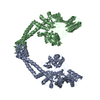
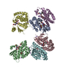
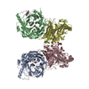
| Title | Crystal Structure of Human Calcineurin in Complex with PVIVIT Peptide | ||||||
 Components Components |
| ||||||
 Keywords Keywords | HYDROLASE/HYDROLASE REGULATOR / Beta-sheet Augmentation / Protein-peptide Complex / HYDROLASE-HYDROLASE REGULATOR COMPLEX | ||||||
| Function / homology |  Function and homology information Function and homology informationnegative regulation of angiotensin-activated signaling pathway / calcium-dependent protein serine/threonine phosphatase regulator activity / regulation of cell proliferation involved in kidney morphogenesis / positive regulation of glomerulus development / negative regulation of calcium ion import across plasma membrane / negative regulation of signaling / calcium-dependent protein serine/threonine phosphatase activity / protein serine/threonine phosphatase complex / positive regulation of saliva secretion / peptidyl-serine dephosphorylation ...negative regulation of angiotensin-activated signaling pathway / calcium-dependent protein serine/threonine phosphatase regulator activity / regulation of cell proliferation involved in kidney morphogenesis / positive regulation of glomerulus development / negative regulation of calcium ion import across plasma membrane / negative regulation of signaling / calcium-dependent protein serine/threonine phosphatase activity / protein serine/threonine phosphatase complex / positive regulation of saliva secretion / peptidyl-serine dephosphorylation / calmodulin-dependent protein phosphatase activity / calcineurin complex / positive regulation of calcium ion-dependent exocytosis of neurotransmitter / positive regulation of connective tissue replacement / positive regulation of calcium ion import across plasma membrane / negative regulation of dendrite morphogenesis / positive regulation of cardiac muscle hypertrophy in response to stress / renal filtration / lung epithelial cell differentiation / calcineurin-NFAT signaling cascade / positive regulation of calcineurin-NFAT signaling cascade / transition between fast and slow fiber / skeletal muscle tissue regeneration / myelination in peripheral nervous system / positive regulation of osteoclast differentiation / cardiac muscle hypertrophy in response to stress / regulation of synaptic vesicle cycle / dephosphorylation / extrinsic component of plasma membrane / branching involved in blood vessel morphogenesis / dendrite morphogenesis / protein dephosphorylation / protein-serine/threonine phosphatase / CLEC7A (Dectin-1) induces NFAT activation / regulation of postsynaptic neurotransmitter receptor internalization / protein serine/threonine phosphatase activity / calcineurin-mediated signaling / parallel fiber to Purkinje cell synapse / positive regulation of activated T cell proliferation / epithelial to mesenchymal transition / Calcineurin activates NFAT / positive regulation of endocytosis / epidermis development / Activation of BAD and translocation to mitochondria / DARPP-32 events / positive regulation of osteoblast differentiation / multicellular organismal response to stress / phosphatase binding / postsynaptic modulation of chemical synaptic transmission / keratinocyte differentiation / skeletal muscle fiber development / FCERI mediated Ca+2 mobilization / positive regulation of cell adhesion / T cell activation / hippocampal mossy fiber to CA3 synapse / excitatory postsynaptic potential / wound healing / G1/S transition of mitotic cell cycle / response to calcium ion / sarcolemma / modulation of chemical synaptic transmission / Schaffer collateral - CA1 synapse / Z disc / protein import into nucleus / calcium ion transport / heart development / ATPase binding / Ca2+ pathway / dendritic spine / calmodulin binding / protein dimerization activity / postsynapse / positive regulation of cell migration / protein domain specific binding / negative regulation of gene expression / calcium ion binding / positive regulation of gene expression / glutamatergic synapse / enzyme binding / positive regulation of transcription by RNA polymerase II / mitochondrion / nucleoplasm / plasma membrane / cytoplasm / cytosol Similarity search - Function | ||||||
| Biological species |  Homo sapiens (human) Homo sapiens (human) | ||||||
| Method |  X-RAY DIFFRACTION / SYNCHROTRON / MOLECULAR REPLACEMENT / Resolution: 2.3 Å X-RAY DIFFRACTION / SYNCHROTRON / MOLECULAR REPLACEMENT / Resolution: 2.3 Å | ||||||
 Authors Authors | Li, H. / Zhang, L. / Rao, A. / Harrison, S.C. / Hogan, P.G. | ||||||
 Citation Citation | Journal: J.Mol.Biol. / Year: 2007 Title: Structure of calcineurin in complex with PVIVIT peptide: Portrait of a low-affinity signalling interaction Authors: Li, H. / Zhang, L. / Rao, A. / Harrison, S.C. / Hogan, P.G. #1: Journal: Nature / Year: 1995Title: Crystal Structures of Human Calcineurin and the Human FKBP12-FK506-Calcineurin Complex Authors: Kissinger, C.R. / Parge, H.E. / Knighton, D.R. / Lewis, C.T. / Pelletier, L.A. / Tempczyk, A. / Kalish, V.J. / Tucker, K.D. / Showalter, R.E. / Moomaw, E.W. / Gastinel, L.N. / Habuka, N. / ...Authors: Kissinger, C.R. / Parge, H.E. / Knighton, D.R. / Lewis, C.T. / Pelletier, L.A. / Tempczyk, A. / Kalish, V.J. / Tucker, K.D. / Showalter, R.E. / Moomaw, E.W. / Gastinel, L.N. / Habuka, N. / Chen, X. / Maldonado, F. / Barker, J.E. #2: Journal: Cell(Cambridge,Mass.) / Year: 1995Title: X-ray Structure of Calcineurin Inhibited by the Immunophilin-immunosuppressant FKBP12-FK506 Complex Authors: Griffith, J.P. / Kim, J.L. / Kim, E.E. / Sintchak, M.D. / Thomson, J.A. / Fitzgibbon, M.J. / Fleming, M.A. / Caron, P.R. / Hsiao, K. / Navia, M.A. #3: Journal: Science / Year: 1999Title: Affinity-driven Peptide Selection of an NFAT Inhibitor More Selective Than Cyclosporin A Authors: Aramburu, J. / Yaffe, M.B. / Lopez-Rodriguez, C. / Cantley, L.C. / Hogan, P.G. / Rao, A. #4: Journal: J.Mol.Biol. / Year: 2004Title: Structural Delineation of the Calcineurin-NFAT Interaction and Its Parallels to PP1 Targeting Interactions Authors: Li, H. / Rao, A. / Hogan, P.G. | ||||||
| History |
|
- Structure visualization
| Structure viewer | Molecule: MolmilJmol/JSmol |
|---|
- Downloads & links
-Download
| PDBx/mmCIF format | 2p6b.cif.gz | 236.8 KB | Display | PDBx/mmCIF format |
|---|---|---|---|---|
| PDB format | pdb2p6b.ent.gz | 185.3 KB | Display | PDB format |
| PDBx/mmJSON format | 2p6b.json.gz | Tree view | PDBx/mmJSON format | |
| Others |  Other downloads Other downloads |
-Validation report
| Arichive directory | https://data.pdbj.org/pub/pdb/validation_reports/p6/2p6bftp://data.pdbj.org/pub/pdb/validation_reports/p6/2p6b | HTTPS FTP |
|---|
-Related structure data
| Related structure data | 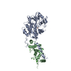 1auiS S: Starting model for refinement |
|---|---|
| Similar structure data |


-Links
 PDBj
PDBj


- Assembly
| Deposited unit | 
| ||||||||
|---|---|---|---|---|---|---|---|---|---|
| 1 |
| ||||||||
| Unit cell |
| ||||||||
| Details | Although the asymmetric unit contains two calcineurin AB heterodimers, the functional unit in solution is a single AB heterodimer |
-Components
-Protein/peptide , 1 types, 1 molecules E
| #1: Protein/peptide | Mass: 1481.673 Da / Num. of mol.: 1 / Fragment: Residues 3-16 / Source method: obtained synthetically / Details: Synthetic Peptide |
|---|
-Protein , 2 types, 4 molecules ACBD
| #2: Protein | Mass: 43991.188 Da / Num. of mol.: 2 / Fragment: Residues 1-381 Source method: isolated from a genetically manipulated source Source: (gene. exp.) Homo sapiens (human) / Gene: PPP3CA, CALNA, CNA / Plasmid: pET15b / Species (production host): Escherichia coli / Production host:  #3: Protein | Mass: 17755.174 Da / Num. of mol.: 2 / Fragment: Residues 16-170 Source method: isolated from a genetically manipulated source Source: (gene. exp.) Homo sapiens (human) / Gene: PPP3R1 / Plasmid: pET15b / Species (production host): Escherichia coli / Production host: |
|---|
-Non-polymers , 5 types, 578 molecules 


| #4: Chemical |  Mass: 65.409 Da / Num. of mol.: 2 / Source method: obtained synthetically / Formula: Zn Mass: 65.409 Da / Num. of mol.: 2 / Source method: obtained synthetically / Formula: Zn#5: Chemical |  Mass: 55.845 Da / Num. of mol.: 2 / Source method: obtained synthetically / Formula: Fe Mass: 55.845 Da / Num. of mol.: 2 / Source method: obtained synthetically / Formula: Fe#6: Chemical |  Mass: 94.971 Da / Num. of mol.: 2 / Source method: obtained synthetically / Formula: PO4 Mass: 94.971 Da / Num. of mol.: 2 / Source method: obtained synthetically / Formula: PO4#7: Chemical | ChemComp-CA /  Mass: 40.078 Da / Num. of mol.: 8 / Source method: obtained synthetically / Formula: Ca Mass: 40.078 Da / Num. of mol.: 8 / Source method: obtained synthetically / Formula: Ca#8: Water | ChemComp-HOH / | Mass: 18.015 Da / Num. of mol.: 564 / Source method: isolated from a natural source / Formula: H2O |
|---|
-Details
| Has protein modification | Y |
|---|
-Experimental details
-Experiment
| Experiment | Method: X-RAY DIFFRACTION / Number of used crystals: 1 |
|---|
- Sample preparation
| Crystal | Density Matthews: 2.42 Å3/Da / Density % sol: 49.18 % |
|---|---|
| Crystal grow | Temperature: 295 K / Method: vapor diffusion, hanging drop / pH: 8 Details: 12% PEG 4000, 0.1M calcium chloride, 0.1M TES, 0.001M DTT, pH 8.0, VAPOR DIFFUSION, HANGING DROP, temperature 295K |
-Data collection
| Diffraction | Mean temperature: 100 K |
|---|---|
| Diffraction source | Source: SYNCHROTRON / Site: APS  / Beamline: 19-ID / Wavelength: 0.9789 Å / Beamline: 19-ID / Wavelength: 0.9789 Å |
| Detector | Type: ADSC QUANTUM 315 / Detector: CCD / Date: Feb 24, 2006 Details: Rosenbaum-Rock high-resolution double-crystal monochromator. LN2 cooled first crystal, sagittal focusing 2nd crystal, Rosenbaum-Rock vertical focusing mirror, beam defining slits |
| Radiation | Monochromator: Si 111 / Protocol: SINGLE WAVELENGTH / Monochromatic (M) / Laue (L): M / Scattering type: x-ray |
| Radiation wavelength | Wavelength: 0.9789 Å / Relative weight: 1 |
| Reflection | Resolution: 2.2→50 Å / Num. obs: 60710 / % possible obs: 96.2 % / Redundancy: 4.7 % / Rmerge(I) obs: 0.104 / Χ2: 1.563 / Net I/σ(I): 8.2 |
| Reflection shell | Resolution: 2.2→2.28 Å / Redundancy: 1.9 % / Rmerge(I) obs: 0.529 / Num. unique all: 4593 / Χ2: 1.279 / % possible all: 73.9 |
-Phasing
| Phasing MR |
|
|---|
- Processing
| Software |
| ||||||||||||||||||||||||||||||||||||
|---|---|---|---|---|---|---|---|---|---|---|---|---|---|---|---|---|---|---|---|---|---|---|---|---|---|---|---|---|---|---|---|---|---|---|---|---|---|
| Refinement | Method to determine structure: MOLECULAR REPLACEMENT Starting model: PDB Entry 1AUI Resolution: 2.3→50 Å / Cross valid method: THROUGHOUT / σ(F): 0 / Stereochemistry target values: Engh & Huber
| ||||||||||||||||||||||||||||||||||||
| Solvent computation | Bsol: 36.056 Å2 | ||||||||||||||||||||||||||||||||||||
| Displacement parameters | Biso mean: 37.883 Å2
| ||||||||||||||||||||||||||||||||||||
| Refinement step | Cycle: LAST / Resolution: 2.3→50 Å
| ||||||||||||||||||||||||||||||||||||
| Refine LS restraints |
| ||||||||||||||||||||||||||||||||||||
| LS refinement shell | Highest resolution: 2.3 Å
| ||||||||||||||||||||||||||||||||||||
| Xplor file |
|
