 Movie
Movie Controller

[English] 日本語
 Yorodumi
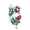
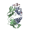
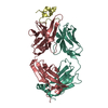
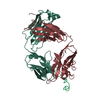
- PDB-2osl: Crystal structure of Rituximab Fab in complex with an epitope peptide -
+ Open data

- Basic information
| Entry | Database: PDB / ID: 2osl | ||||||
|---|---|---|---|---|---|---|---|
| Title | Crystal structure of Rituximab Fab in complex with an epitope peptide | ||||||
 Components Components |
| ||||||
 Keywords Keywords |  IMMUNE SYSTEM / Fab-peptide complex / Rituximab / chimeric antibody IMMUNE SYSTEM / Fab-peptide complex / Rituximab / chimeric antibody | ||||||
| Function / homology |  Function and homology information Function and homology informationstore-operated calcium entry / positive regulation of calcium ion import across plasma membrane / calcium ion import into cytosol / epidermal growth factor receptor binding / B cell activation / B cell proliferation / plasma membrane raft / immunoglobulin binding / humoral immune response / B cell differentiation ...store-operated calcium entry / positive regulation of calcium ion import across plasma membrane / calcium ion import into cytosol / epidermal growth factor receptor binding / B cell activation / B cell proliferation / plasma membrane raft / immunoglobulin binding / humoral immune response / B cell differentiation / response to bacterium / protein tetramerization / B cell receptor signaling pathway / MHC class II protein complex binding / cell surface receptor signaling pathway / external side of plasma membrane / cell surface / extracellular space / extracellular exosome / nucleoplasm / identical protein binding / plasma membraneSimilarity search - Function | ||||||
| Biological species |  Mus musculus (house mouse) Mus musculus (house mouse) Homo sapiens (human) Homo sapiens (human) | ||||||
| Method | X-RAY DIFFRACTION / SYNCHROTRON / MOLECULAR REPLACEMENT / Resolution: 2.6 Å | ||||||
 Authors Authors | Du, J. / Zhong, C. / Ding, J. | ||||||
 Citation Citation | Journal: J.Biol.Chem. / Year: 2007 Title: Structural basis for recognition of CD20 by therapeutic antibody Rituximab Authors: Du, J. / Wang, H. / Zhong, C. / Peng, B. / Zhang, M. / Li, B. / Huo, S. / Guo, Y. / Ding, J. | ||||||
| History |
| ||||||
| Remark 999 | SEQUENCE Sequence database references for chain L, B and chain H, A do not currently exist. |
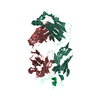
- Structure visualization
| Structure viewer | Molecule: MolmilJmol/JSmol |
|---|
- Downloads & links
-Download
| PDBx/mmCIF format | 2osl.cif.gz | 185.5 KB | Display | PDBx/mmCIF format |
|---|---|---|---|---|
| PDB format | pdb2osl.ent.gz | 148.2 KB | Display | PDB format |
| PDBx/mmJSON format | 2osl.json.gz | Tree view | PDBx/mmJSON format | |
| Others |  Other downloads Other downloads |
-Validation report
| Arichive directory | https://data.pdbj.org/pub/pdb/validation_reports/os/2oslftp://data.pdbj.org/pub/pdb/validation_reports/os/2osl | HTTPS FTP |
|---|
-Related structure data
| Related structure data | 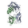 1ad0S S: Starting model for refinement |
|---|---|
| Similar structure data |


-Links
 PDBj
PDBj

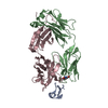
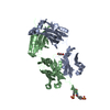
- Assembly
| Deposited unit | 
| ||||||||
|---|---|---|---|---|---|---|---|---|---|
| 1 | 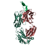
| ||||||||
| 2 | 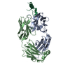
| ||||||||
| Unit cell |
| ||||||||
| Details | the Biological Assembly is monomer. |
-Components
| #1: Antibody | Mass: 23078.623 Da / Num. of mol.: 2 Source method: isolated from a genetically manipulated source Details: The protein was produced as a chimeric fab fragment. residues 1-106 is from murine and 107-213 is from human. Source: (gene. exp.) Mus musculus (house mouse), (gene. exp.) Homo sapiens (human)Species: , / Description: The antibody was purchased from Roche. / Production host: synthetic construct (others) #2: Antibody | Mass: 23733.541 Da / Num. of mol.: 2 Source method: isolated from a genetically manipulated source Details: The protein was produced as a chimeric fab fragment. residues 1-121 is from murine and 122-224 is from human. Source: (gene. exp.) Mus musculus (house mouse), (gene. exp.) Homo sapiens (human)Species: , / Description: The antibody was purchased from Roche. / Production host: synthetic construct (others) #3: Protein/peptide | Mass: 2852.051 Da / Num. of mol.: 2 / Fragment: epitope peptide / Source method: obtained synthetically Details: the epitope peptide was synthesized at Shanghai Science Peptide Biological Technology Source: (synth.) Homo sapiens (human) / References: UniProt: P11836#4: Water | ChemComp-HOH / | Water Mass: 18.015 Da / Num. of mol.: 212 / Source method: isolated from a natural source / Formula: H2O Mass: 18.015 Da / Num. of mol.: 212 / Source method: isolated from a natural source / Formula: H2O |
|---|
-Experimental details
-Experiment
| Experiment | Method: X-RAY DIFFRACTION / Number of used crystals: 1 |
|---|
- Sample preparation
| Crystal | Density Matthews: 2.57 Å3/Da / Density % sol: 52.14 % |
|---|---|
| Crystal grow | Temperature: 277 K / Method: vapor diffusion, hanging drop / pH: 6.5 Details: 0.2M calcium acetate, 0.1M sodium cacodylate, 18% PEG8000, pH 6.5, VAPOR DIFFUSION, HANGING DROP, temperature 277K |
-Data collection
| Diffraction | Mean temperature: 100 K |
|---|---|
| Diffraction source | Source: SYNCHROTRON / Site: Photon Factory  / Beamline: AR-NW12A / Wavelength: 1 Å / Beamline: AR-NW12A / Wavelength: 1 Å |
| Detector | Type: ADSC QUANTUM 210 / Detector: CCD / Date: Nov 20, 2006 |
| Radiation | Protocol: SINGLE WAVELENGTH / Monochromatic (M) / Laue (L): M / Scattering type: x-ray |
| Radiation wavelength | Wavelength: 1 Å / Relative weight: 1 |
| Reflection | Resolution: 2.6→50 Å / Num. all: 32638 / Num. obs: 29570 / % possible obs: 90.6 % / Redundancy: 4.1 % / Biso Wilson estimate: 62.749 Å2 / Rmerge(I) obs: 0.076 / Rsym value: 0.076 / Χ2: 0.892 / Net I/σ(I): 7.9 |
| Reflection shell | Resolution: 2.6→2.69 Å / Redundancy: 3.7 % / Rmerge(I) obs: 0.434 / Mean I/σ(I) obs: 2 / Num. unique all: 2958 / Rsym value: 0.434 / Χ2: 0.492 / % possible all: 91.5 |
- Processing
| Software |
| ||||||||||||||||||||||||||||||||||||
|---|---|---|---|---|---|---|---|---|---|---|---|---|---|---|---|---|---|---|---|---|---|---|---|---|---|---|---|---|---|---|---|---|---|---|---|---|---|
| Refinement | Method to determine structure: MOLECULAR REPLACEMENT Starting model: PDB ENTRY 1AD0 Resolution: 2.6→43.97 Å / Rfactor Rfree error: 0.008 / Data cutoff high absF: 1491027.625 / Data cutoff low absF: 0 / Isotropic thermal model: RESTRAINED / Cross valid method: THROUGHOUT / σ(F): 0 / σ(I): 0
| ||||||||||||||||||||||||||||||||||||
| Solvent computation | Solvent model: FLAT MODEL / Bsol: 35.903 Å2 / ksol: 0.341 e/Å3 | ||||||||||||||||||||||||||||||||||||
| Displacement parameters | Biso mean: 49 Å2
| ||||||||||||||||||||||||||||||||||||
| Refine analyze |
| ||||||||||||||||||||||||||||||||||||
| Refinement step | Cycle: LAST / Resolution: 2.6→43.97 Å
| ||||||||||||||||||||||||||||||||||||
| Refine LS restraints |
| ||||||||||||||||||||||||||||||||||||
| LS refinement shell | Resolution: 2.6→2.76 Å / Rfactor Rfree error: 0.028 / Total num. of bins used: 6
| ||||||||||||||||||||||||||||||||||||
| Xplor file |
|
