 Movie
Movie Controller


+ Open data

- Basic information
| Entry | Database: PDB / ID: 2jk4 | ||||||
|---|---|---|---|---|---|---|---|
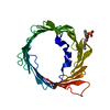
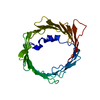
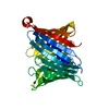
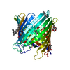
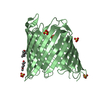
| Title | Structure of the human voltage-dependent anion channel | ||||||
 Components Components | VOLTAGE-DEPENDENT ANION-SELECTIVE CHANNEL PROTEIN 1 | ||||||
 Keywords Keywords | TRANSPORT / VDAC /  PORIN / MEMBRANE / APOPTOSIS / MITOCHONDRION OUTER MEMBRANE / MITOCHONDRIAL OUTER MEMBRANE / MEMBRANE PROTEIN / HOST-VIRUS INTERACTION / ION TRANSPORT / TRANSMEMBRANE / PHOSPHOPROTEIN / ACETYLATION / MITOCHONDRION / CELL MEMBRANE PORIN / MEMBRANE / APOPTOSIS / MITOCHONDRION OUTER MEMBRANE / MITOCHONDRIAL OUTER MEMBRANE / MEMBRANE PROTEIN / HOST-VIRUS INTERACTION / ION TRANSPORT / TRANSMEMBRANE / PHOSPHOPROTEIN / ACETYLATION / MITOCHONDRION / CELL MEMBRANE | ||||||
| Function / homology |  Function and homology information Function and homology informationnegative regulation of calcium import into the mitochondrion / positive regulation of parkin-mediated stimulation of mitophagy in response to mitochondrial depolarization / voltage-gated monoatomic anion channel activity / neuron-neuron synaptic transmission / Mitochondrial calcium ion transport / ceramide binding / regulation of autophagy of mitochondrion / mitochondrial permeability transition pore complex / Pyruvate metabolism / Mitochondrial protein import ...negative regulation of calcium import into the mitochondrion / positive regulation of parkin-mediated stimulation of mitophagy in response to mitochondrial depolarization / voltage-gated monoatomic anion channel activity / neuron-neuron synaptic transmission / Mitochondrial calcium ion transport / ceramide binding / regulation of autophagy of mitochondrion / mitochondrial permeability transition pore complex / Pyruvate metabolism / Mitochondrial protein import / phosphatidylcholine binding / pyruvate metabolic process / oxysterol binding / monoatomic anion transport / cholesterol binding / porin activity / mitochondrial nucleoid / pore complex / negative regulation of reactive oxygen species metabolic process / behavioral fear response / epithelial cell differentiation / PINK1-PRKN Mediated Mitophagy / learning / mitochondrial membrane / mitochondrial outer membrane / transmembrane transporter binding / Ub-specific processing proteases / membrane raft / synapse / apoptotic process / negative regulation of apoptotic process / protein kinase binding / mitochondrion / extracellular exosome / membrane / identical protein binding / nucleus / plasma membraneSimilarity search - Function | ||||||
| Biological species |  HOMO SAPIENS (human) HOMO SAPIENS (human) | ||||||
| Method | X-RAY DIFFRACTION / SYNCHROTRON / OTHER / Resolution: 4.1 Å | ||||||
 Authors Authors | Bayrhuber, M. / Meins, T. / Habeck, M. / Becker, S. / Giller, K. / Villinger, S. / Vonrhein, C. / Griesinger, C. / Zweckstetter, M. / Zeth, K. | ||||||
 Citation Citation | Journal: Proc.Natl.Acad.Sci.USA / Year: 2008 Title: Structure of the Human Voltage-Dependent Anion Channel. Authors: Bayrhuber, M. / Meins, T. / Habeck, M. / Becker, S. / Giller, K. / Villinger, S. / Vonrhein, C. / Griesinger, C. / Zweckstetter, M. / Zeth, K. | ||||||
| History |
| ||||||
| Remark 700 | SHEET DETERMINATION METHOD: AUTHOR PROVIDED. |
- Structure visualization
| Structure viewer | Molecule: MolmilJmol/JSmol |
|---|
- Downloads & links
-Download
| PDBx/mmCIF format | 2jk4.cif.gz | 55.9 KB | Display | PDBx/mmCIF format |
|---|---|---|---|---|
| PDB format | pdb2jk4.ent.gz | 42.3 KB | Display | PDB format |
| PDBx/mmJSON format | 2jk4.json.gz | Tree view | PDBx/mmJSON format | |
| Others |  Other downloads Other downloads |
-Validation report
| Arichive directory | https://data.pdbj.org/pub/pdb/validation_reports/jk/2jk4ftp://data.pdbj.org/pub/pdb/validation_reports/jk/2jk4 | HTTPS FTP |
|---|
-Related structure data
| Similar structure data |
|---|


-Links
 PDBj
PDBj

- Assembly
| Deposited unit | 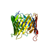
| ||||||||
|---|---|---|---|---|---|---|---|---|---|
| 1 |
| ||||||||
| Unit cell |
|
-Components
| #1: Protein | Mass: 32181.988 Da / Num. of mol.: 1 / Fragment: RESIDUES 2-283 Source method: isolated from a genetically manipulated source Source: (gene. exp.) HOMO SAPIENS (human) / Production host:  ESCHERICHIA COLI (E. coli) / References: UniProt: P21796 ESCHERICHIA COLI (E. coli) / References: UniProt: P21796 |
|---|
-Experimental details
-Experiment
| Experiment | Method: X-RAY DIFFRACTION / Number of used crystals: 1 |
|---|
- Sample preparation
| Crystal | Density Matthews: 4.5 Å3/Da / Density % sol: 73 % / Description: NONE |
|---|---|
| Crystal grow | Details: 0.2 M MAGNESIUM CHLORIDE HEXAHYDRATE, 0.1 M HEPES SODIUM PH 7.5, 30% V/V POLYETHYLENE GLYCOL 400 |
-Data collection
| Diffraction | Mean temperature: 100 K |
|---|---|
| Diffraction source | Source: SYNCHROTRON / Site: ESRF  / Beamline: ID29 / Wavelength: 0.9537 / Beamline: ID29 / Wavelength: 0.9537 |
| Detector | Type: ADSC CCD / Detector: CCD / Date: Feb 24, 2006 |
| Radiation | Protocol: SINGLE WAVELENGTH / Monochromatic (M) / Laue (L): M / Scattering type: x-ray |
| Radiation wavelength | Wavelength: 0.9537 Å / Relative weight: 1 |
| Reflection | Resolution: 4.1→43 Å / Num. obs: 4887 / % possible obs: 99.5 % / Observed criterion σ(I): 2 / Redundancy: 16.8 % / Biso Wilson estimate: 175.334 Å2 / Rmerge(I) obs: 0.1 / Net I/σ(I): 16.4 |
| Reflection shell | Resolution: 4.1→4.3 Å / Redundancy: 8.3 % / Rmerge(I) obs: 0.54 / Mean I/σ(I) obs: 3.5 / % possible all: 99.1 |
- Processing
| Software |
| ||||||||||||||||||||
|---|---|---|---|---|---|---|---|---|---|---|---|---|---|---|---|---|---|---|---|---|---|
| Refinement | Method to determine structure: OTHER Starting model: NONE Resolution: 4.1→25 Å / Cross valid method: THROUGHOUT / σ(F): 0 / Details: HYDROGENS HAVE BEEN ADDED IN THE RIDING POSITIONS.
| ||||||||||||||||||||
| Displacement parameters | Biso mean: 179.12 Å2
| ||||||||||||||||||||
| Refinement step | Cycle: LAST / Resolution: 4.1→25 Å
| ||||||||||||||||||||
| LS refinement shell | Resolution: 4.1→4.35 Å / Total num. of bins used: 9
|
