National Institutes of Health/National Institute of General Medical Sciences (NIH/NIGMS)
P41GM136508
United States
National Institutes of Health/National Institute of General Medical Sciences (NIH/NIGMS)
R35GM135175
United States
Citation
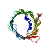
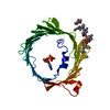
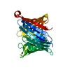
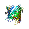
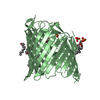
Journal: Proc Natl Acad Sci U S A / Year: 2020 Title: MicroED structure of lipid-embedded mammalian mitochondrial voltage-dependent anion channel. Authors: Michael W Martynowycz / Farha Khan / Johan Hattne / Jeff Abramson / Tamir Gonen / Abstract: A structure of the murine voltage-dependent anion channel (VDAC) was determined by microcrystal electron diffraction (MicroED). Microcrystals of an essential mutant of VDAC grew in a viscous bicelle ...A structure of the murine voltage-dependent anion channel (VDAC) was determined by microcrystal electron diffraction (MicroED). Microcrystals of an essential mutant of VDAC grew in a viscous bicelle suspension, making it unsuitable for conventional X-ray crystallography. Thin, plate-like crystals were identified using scanning-electron microscopy (SEM). Crystals were milled into thin lamellae using a focused-ion beam (FIB). MicroED data were collected from three crystal lamellae and merged for completeness. The refined structure revealed unmodeled densities between protein monomers, indicative of lipids that likely mediate contacts between the proteins in the crystal. This body of work demonstrates the effectiveness of milling membrane protein microcrystals grown in viscous media using a focused ion beam for subsequent structure determination by MicroED. This approach is well suited for samples that are intractable by X-ray crystallography. To our knowledge, the presented structure is a previously undescribed mutant of the membrane protein VDAC, crystallized in a lipid bicelle matrix and solved by MicroED.
Voltage-dependentanion-selectivechannelprotein1 / mVDAC1 / Outer mitochondrial membrane protein porin 1 / Plasmalemmal porin / Voltage-dependent ...mVDAC1 / Outer mitochondrial membrane protein porin 1 / Plasmalemmal porin / Voltage-dependent anion-selective channel protein 5 / mVDAC5
Mass: 32195.879 Da / Num. of mol.: 1 / Mutation: K12E Source method: isolated from a genetically manipulated source Source: (gene. exp.) Mus musculus (house mouse) / Gene: Vdac1, Vdac5 / Production host: Escherichia coli (E. coli) / References: UniProt: Q60932
-
Experimental details
-
Experiment
Experiment
Method: ELECTRON CRYSTALLOGRAPHY
EM experiment
Aggregation state: 3D ARRAY / 3D reconstruction method: electron crystallography
Cryogen: NITROGEN / Specimen holder model: FEI TITAN KRIOS AUTOGRID HOLDER / Temperature (max): 90 K / Temperature (min): 77 K
Image recording
Average exposure time: 5 sec. / Electron dose: 0.01 e/Å2 / Film or detector model: FEI CETA (4k x 4k) / Num. of diffraction images: 180 / Num. of grids imaged: 2 / Num. of real images: 150
Image scans
Sampling size: 28 µm / Width: 2048 / Height: 2048
EM diffraction
Camera length: 3000 mm / Tilt angle list: -60,68
EM diffraction shell
Resolution: 3.232→3.12 Å / Fourier space coverage: 57.5 % / Multiplicity: 6.2 / Num. of structure factors: 565 / Phase residual: 27 °
EM diffraction stats
Fourier space coverage: 76.29 % / High resolution: 3.12 Å / Num. of intensities measured: 32723 / Num. of structure factors: 5410 / Phase error: 25 ° / Phase residual: 25 ° / Phase error rejection criteria: none used / Rmerge: 0.47 / Rsym: 0.22
Reflection
Biso Wilson estimate: 93.7 Å2
-
Processing
Software
Name
Version
Classification
NB
phenix.refine
1.18.2_3874+SVN
refinement
PHENIX
1.18.2_3874+SVN
refinement
EM software
ID
Name
Version
Category
Details (eV)
1
EPU
imageacquisition
EPU-D
6
PHENIX
1.18.2
modelfitting
Phaser
8
PHENIX
1.18.2
modelrefinement
phenix.refine
9
PHENIX
molecularreplacement
Phaser
12
AIMLESS
crystallographymerging
13
PHENIX
3Dreconstruction
Image processing
Details: CetaD 2x binned
EM 3D crystal entity
∠α: 90 ° / ∠β: 99.44 ° / ∠γ: 90 ° / A: 98.9 Å / B: 58.21 Å / C: 69.54 Å / Space group name: C121 / Space group num: 5
CTF correction
Type: NONE
3D reconstruction
Resolution: 3.12 Å / Resolution method: DIFFRACTION PATTERN/LAYERLINES / Algorithm: FOURIER SPACE / Symmetry type: 3D CRYSTAL
Atomic model building
B value: 90 / Protocol: RIGID BODY FIT / Space: RECIPROCAL / Target criteria: Maximum liklihood Details: Standard refinement with electron scattering factors
In the structure databanks used in Yorodumi, some data are registered as the other names, "COVID-19 virus" and "2019-nCoV". Here are the details of the virus and the list of structure data.
Jan 31, 2019. EMDB accession codes are about to change! (news from PDBe EMDB page)
EMDB accession codes are about to change! (news from PDBe EMDB page)
The allocation of 4 digits for EMDB accession codes will soon come to an end. Whilst these codes will remain in use, new EMDB accession codes will include an additional digit and will expand incrementally as the available range of codes is exhausted. The current 4-digit format prefixed with “EMD-” (i.e. EMD-XXXX) will advance to a 5-digit format (i.e. EMD-XXXXX), and so on. It is currently estimated that the 4-digit codes will be depleted around Spring 2019, at which point the 5-digit format will come into force.
The EM Navigator/Yorodumi systems omit the EMD- prefix.
Related info.:Q: What is EMD? / ID/Accession-code notation in Yorodumi/EM Navigator
Yorodumi is a browser for structure data from EMDB, PDB, SASBDB, etc.
This page is also the successor to EM Navigator detail page, and also detail information page/front-end page for Omokage search.
The word "yorodu" (or yorozu) is an old Japanese word meaning "ten thousand". "mi" (miru) is to see.
Related info.:EMDB / PDB / SASBDB / Comparison of 3 databanks / Yorodumi Search / Aug 31, 2016. New EM Navigator & Yorodumi / Yorodumi Papers / Jmol/JSmol / Function and homology information / Changes in new EM Navigator and Yorodumi
 Movie
Movie Controller


 Open data
 Basic information
 Components
 Keywords
 Function and homology information
 Authors
 United States, 3items
  Citation
 Structure visualization
 Downloads & links
 Other downloads


 PDBj
PDBj

 Assembly
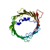

 Sample preparation
 FIELD EMISSION GUN / Accelerating voltage: 300 kV / Illumination mode: FLOOD BEAM
 Processing