 Movie
Movie Controller


+ Open data

- Basic information
| Entry | Database: PDB / ID: 2jia | ||||||
|---|---|---|---|---|---|---|---|
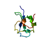
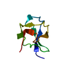
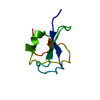
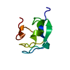
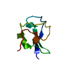
| Title | TYPE III ANTIFREEZE PROTEIN ISOFORM HPLC 12 K61I | ||||||
 Components Components | PROTEIN (ANTIFREEZE PROTEIN TYPE III) | ||||||
 Keywords Keywords | ANTIFREEZE PROTEIN / MUTANT / ICE BINDING PROTEIN / THERMAL HYSTERESIS PROTEIN | ||||||
| Function / homology |  Function and homology information Function and homology information | ||||||
| Biological species |  Macrozoarces americanus (ocean pout) Macrozoarces americanus (ocean pout) | ||||||
| Method |  X-RAY DIFFRACTION / OTHER / Resolution: 1.6 Å X-RAY DIFFRACTION / OTHER / Resolution: 1.6 Å | ||||||
 Authors Authors | Graether, S.P. / Deluca, C.I. / Baardsnes, J. / Hill, G.A. / Davies, P.L. / Jia, Z. | ||||||
 Citation Citation | Journal: J.Biol.Chem. / Year: 1999 Title: Quantitative and qualitative analysis of type III antifreeze protein structure and function. Authors: Graether, S.P. / DeLuca, C.I. / Baardsnes, J. / Hill, G.A. / Davies, P.L. / Jia, Z. #1: Journal: J.Mol.Biol. / Year: 1998Title: The Effects of Steric Mutations on the Structure of Type III Antifreeze Protein and its Interaction with Ice Authors: Deluca, C.I. / Davies, P.L. / Ye, Q. / Jia, Z. #2: Journal: Nature / Year: 1996Title: Structural Basis for the Binding of a Globular Antifreeze Protein to Ice Authors: Jia, Z. / Deluca, C.I. / Chao, H. / Davies, P.L. #3: Journal: Protein Sci. / Year: 1995Title: Crystallization and Preliminary X-Ray Crystallographic Studies on Type III Antifreeze Protein Authors: Jia, Z. / Deluca, C.I. / Davies, P.L. #4: Journal: Protein Sci. / Year: 1993Title: Use of Proline Mutants to Help Solve the NMR Solution Structure of Type III Antifreeze Protein Authors: Chao, H. / Davies, P.L. / Sykes, B.D. / Sonnichsen, F.D. #5: Journal: J.Biol.Chem. / Year: 1988Title: Multiple Genes Provide the Basis for Antifreeze Protein Diversity and Dosage in the Ocean Pout, Macrozoarces Americanus Authors: Hew, C.L. / Wang, N.C. / Joshi, S. / Fletcher, G.L. / Scott, G.K. / Hayes, P.H. / Buettner, B. / Davies, P.L. | ||||||
| History |
|
- Structure visualization
| Structure viewer | Molecule: MolmilJmol/JSmol |
|---|
- Downloads & links
-Download
| PDBx/mmCIF format | 2jia.cif.gz | 27.9 KB | Display | PDBx/mmCIF format |
|---|---|---|---|---|
| PDB format | pdb2jia.ent.gz | 18.6 KB | Display | PDB format |
| PDBx/mmJSON format | 2jia.json.gz | Tree view | PDBx/mmJSON format | |
| Others |  Other downloads Other downloads |
-Validation report
| Arichive directory | https://data.pdbj.org/pub/pdb/validation_reports/ji/2jiaftp://data.pdbj.org/pub/pdb/validation_reports/ji/2jia | HTTPS FTP |
|---|
-Related structure data
| Related structure data | 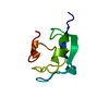 1b7iC 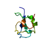 1b7jC  1b7kC  1eklC  1jabC 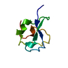 1msjC  2ameC 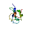 2msjC 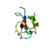 2spgC 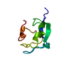 3ameC 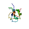 4ameC 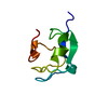 6ameC 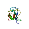 7ameC 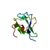 8ameC 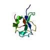 8msiC 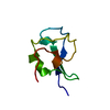 9ameC  9msiC 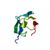 1msiS S: Starting model for refinement C: citing same article ( |
|---|---|
| Similar structure data |


-Links
 PDBj
PDBj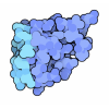
- Assembly
| Deposited unit | 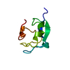
| ||||||||
|---|---|---|---|---|---|---|---|---|---|
| 1 |
| ||||||||
| Unit cell |
|
-Components
| #1: Protein | Mass: 6963.210 Da / Num. of mol.: 1 / Mutation: K61I, P64A, P65A Source method: isolated from a genetically manipulated source Source: (gene. exp.) Macrozoarces americanus (ocean pout) / Plasmid: PT7-7F / Production host:  |
|---|---|
| #2: Water | ChemComp-HOH /  Mass: 18.015 Da / Num. of mol.: 52 / Source method: isolated from a natural source / Formula: H2O Mass: 18.015 Da / Num. of mol.: 52 / Source method: isolated from a natural source / Formula: H2O |
-Experimental details
-Experiment
| Experiment | Method: X-RAY DIFFRACTION / Number of used crystals: 1 |
|---|
- Sample preparation
| Crystal | Density Matthews: 2.16 Å3/Da / Density % sol: 43.08 % | ||||||||||||||||||||||||||||||
|---|---|---|---|---|---|---|---|---|---|---|---|---|---|---|---|---|---|---|---|---|---|---|---|---|---|---|---|---|---|---|---|
| Crystal grow | pH: 5.5 / Details: pH 5.5 | ||||||||||||||||||||||||||||||
| Crystal grow | *PLUS Temperature: 20 ℃ / Method: vapor diffusion, hanging drop / Details: Jia, Z., (1995) Protein Sci., 4, 1236. / PH range low: 4.5 / PH range high: 4 | ||||||||||||||||||||||||||||||
| Components of the solutions | *PLUS
|
-Data collection
| Diffraction | Mean temperature: 298 K |
|---|---|
| Diffraction source | Source: ROTATING ANODE / Type: RIGAKU RU200 / Wavelength: 1.5418 |
| Detector | Type: MAR scanner 300 mm plate / Detector: IMAGE PLATE |
| Radiation | Protocol: SINGLE WAVELENGTH / Monochromatic (M) / Laue (L): M / Scattering type: x-ray |
| Radiation wavelength | Wavelength: 1.5418 Å / Relative weight: 1 |
| Reflection | Resolution: 1.6→50 Å / Num. obs: 8191 / % possible obs: 96.6 % / Observed criterion σ(I): 1 / Redundancy: 5.9 % / Rsym value: 0.075 / Net I/σ(I): 27.6 |
| Reflection shell | Resolution: 1.6→1.67 Å / Redundancy: 4.6 % / Mean I/σ(I) obs: 3.7 / Rsym value: 0.367 / % possible all: 72.8 |
- Processing
| Software |
| ||||||||||||||||||||||||||||||||||||||||||||||||||||||||||||
|---|---|---|---|---|---|---|---|---|---|---|---|---|---|---|---|---|---|---|---|---|---|---|---|---|---|---|---|---|---|---|---|---|---|---|---|---|---|---|---|---|---|---|---|---|---|---|---|---|---|---|---|---|---|---|---|---|---|---|---|---|---|
| Refinement | Method to determine structure: OTHER Starting model: 1MSI Resolution: 1.6→8 Å / Rfactor Rfree error: 0.036 / Cross valid method: FREE R / σ(F): 0
| ||||||||||||||||||||||||||||||||||||||||||||||||||||||||||||
| Displacement parameters | Biso mean: 21.3 Å2 | ||||||||||||||||||||||||||||||||||||||||||||||||||||||||||||
| Refinement step | Cycle: LAST / Resolution: 1.6→8 Å
| ||||||||||||||||||||||||||||||||||||||||||||||||||||||||||||
| Refine LS restraints |
| ||||||||||||||||||||||||||||||||||||||||||||||||||||||||||||
| LS refinement shell | Resolution: 1.6→1.67 Å / Rfactor Rfree error: 0.049
| ||||||||||||||||||||||||||||||||||||||||||||||||||||||||||||
| Xplor file | Serial no: 1 / Param file: PARAM19X.PRO / Topol file: TOPH19X.PRO | ||||||||||||||||||||||||||||||||||||||||||||||||||||||||||||
| Software | *PLUS Name: X-PLOR / Version: 3 / Classification: refinement | ||||||||||||||||||||||||||||||||||||||||||||||||||||||||||||
| Refine LS restraints | *PLUS
|
