| Entry | Database: PDB / ID: 2jak
|
|---|
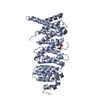
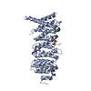
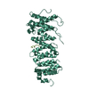
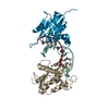
| Title | Human PP2A regulatory subunit B56g |
|---|
 Components Components | SERINE/THREONINE-PROTEIN PHOSPHATASE 2A 56 KDA REGULATORY SUBUNIT GAMMA ISOFORM |
|---|
 Keywords Keywords | NUCLEAR PROTEIN / B56G / PP2A / PPP2R5C / PHOSPHORYLATION / PP2A REGULATORY SUBUNIT |
|---|
| Function / homology |  Function and homology information Function and homology information
protein phosphatase type 2A complex / meiotic sister chromatid cohesion / protein phosphatase regulator activity / APC truncation mutants have impaired AXIN binding / AXIN missense mutants destabilize the destruction complex / Truncations of AMER1 destabilize the destruction complex / Beta-catenin phosphorylation cascade / Signaling by GSK3beta mutants / CTNNB1 S33 mutants aren't phosphorylated / CTNNB1 S37 mutants aren't phosphorylated ...protein phosphatase type 2A complex / meiotic sister chromatid cohesion / protein phosphatase regulator activity / APC truncation mutants have impaired AXIN binding / AXIN missense mutants destabilize the destruction complex / Truncations of AMER1 destabilize the destruction complex / Beta-catenin phosphorylation cascade / Signaling by GSK3beta mutants / CTNNB1 S33 mutants aren't phosphorylated / CTNNB1 S37 mutants aren't phosphorylated / CTNNB1 S45 mutants aren't phosphorylated / CTNNB1 T41 mutants aren't phosphorylated / Co-stimulation by CD28 / Disassembly of the destruction complex and recruitment of AXIN to the membrane / Co-inhibition by CTLA4 / Platelet sensitization by LDL / protein phosphatase activator activity / chromosome, centromeric region / intrinsic apoptotic signaling pathway in response to DNA damage by p53 class mediator / Amplification of signal from unattached kinetochores via a MAD2 inhibitory signal / Mitotic Prometaphase / EML4 and NUDC in mitotic spindle formation / Resolution of Sister Chromatid Cohesion / DNA damage response, signal transduction by p53 class mediator / RAF activation / RHO GTPases Activate Formins / Degradation of beta-catenin by the destruction complex / Negative regulation of MAPK pathway / Separation of Sister Chromatids / Regulation of TP53 Degradation / PI5P, PP2A and IER3 Regulate PI3K/AKT Signaling / proteasome-mediated ubiquitin-dependent protein catabolic process / negative regulation of cell population proliferation / Golgi apparatus / signal transduction / nucleoplasm / nucleus / cytosolSimilarity search - Function Protein phosphatase 2A, regulatory B subunit, B56 / Protein phosphatase 2A regulatory B subunit (B56 family) / Leucine-rich Repeat Variant / Leucine-rich Repeat Variant / Armadillo-like helical / Alpha Horseshoe / Armadillo-type fold / Mainly AlphaSimilarity search - Domain/homology |
|---|
| Biological species |  HOMO SAPIENS (human) HOMO SAPIENS (human) |
|---|
| Method |  X-RAY DIFFRACTION / SYNCHROTRON / MAD / Resolution: 2.6 Å X-RAY DIFFRACTION / SYNCHROTRON / MAD / Resolution: 2.6 Å |
|---|
 Authors Authors | Magnusdottir, A. / Stenmark, P. / Arrowsmith, C. / Berglund, H. / Busam, R. / Collins, R. / Edwards, A. / Ericsson, U.B. / Flodin, S. / Flores, A. ...Magnusdottir, A. / Stenmark, P. / Arrowsmith, C. / Berglund, H. / Busam, R. / Collins, R. / Edwards, A. / Ericsson, U.B. / Flodin, S. / Flores, A. / Graslund, S. / Hammarstrom, M. / Hallberg, B.M. / Holmberg Schiavone, L. / Hogbom, M. / Johansson, I. / Karlberg, T. / Kotenyova, T. / Lundgren, S. / Moche, M. / Nilsson, P. / Nyman, T. / Ogg, D. / Persson, C. / Sagemark, J. / Sundstrom, M. / Uppenberg, J. / Upsten, M. / Thorsell, A.G. / Van Den Berg, S. / Weigelt, J. / Nordlund, P. |
|---|
 Citation Citation | Journal: Proteins / Year: 2009
Title: The Structure of the Pp2A Regulatory Subunit B56 Gamma: The Remaining Piece of the Pp2A Jigsaw Puzzle.
Authors: Magnusdottir, A. / Stenmark, P. / Flodin, S. / Nyman, T. / Kotenyova, T. / Graslund, S. / Ogg, D. / Nordlund, P. |
|---|
| History | | Deposition | Nov 29, 2006 | Deposition site: PDBE / Processing site: PDBE |
|---|
| Revision 1.0 | Dec 4, 2006 | Provider: repository / Type: Initial release |
|---|
| Revision 1.1 | Jul 13, 2011 | Group: Advisory / Version format compliance |
|---|
| Revision 1.2 | May 8, 2024 | Group: Data collection / Database references / Other
Category: chem_comp_atom / chem_comp_bond ...chem_comp_atom / chem_comp_bond / database_2 / pdbx_database_status
Item: _database_2.pdbx_DOI / _database_2.pdbx_database_accession / _pdbx_database_status.status_code_sf |
|---|
|
|---|
 Movie
Movie Controller


 Open data
 Basic information
 Structure visualization
 Downloads & links
 Other downloads
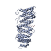


 PDBj
PDBj


 Assembly

 Mass: 18.015 Da / Num. of mol.: 17 / Source method: isolated from a natural source / Formula: H2O
 Sample preparation
 / Beamline: ID14-4 / Wavelength: 1.0689
  Processing