- PDB-2hqw: Crystal Structure of Ca2+/Calmodulin bound to NMDA Receptor NR1C1... -
+
Open data
ID or keywords:
Loading...
-
Basic information
Entry
Database: PDB / ID: 2hqw
Title
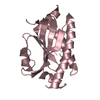
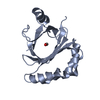
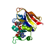
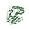
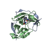
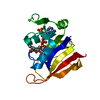
Crystal Structure of Ca2+/Calmodulin bound to NMDA Receptor NR1C1 peptide
Components
Calmodulin
Glutamate NMDA receptor subunit zeta 1
Keywords
METAL BINDING PROTEIN / Calmodulin / EF hand motif / globular complex / ion channel / NR1 / C1 casette / N-methyl-D-aspartate receptor / glutamate / central nervous system / neuronal channel / calcium channel / ER retention signal
Function / homology
Function and homology information
regulation of store-operated calcium channel activity / regulation of high voltage-gated calcium channel activity / excitatory chemical synaptic transmission / : / regulation of response to tumor cell / positive regulation of autophagic cell death / DAPK1-calmodulin complex / pons maturation / regulation of cell communication / positive regulation of Schwann cell migration ...regulation of store-operated calcium channel activity / regulation of high voltage-gated calcium channel activity / excitatory chemical synaptic transmission / : / regulation of response to tumor cell / positive regulation of autophagic cell death / DAPK1-calmodulin complex / pons maturation / regulation of cell communication / positive regulation of Schwann cell migration / : / Synaptic adhesion-like molecules / EPHB-mediated forward signaling / establishment of protein localization to mitochondrial membrane / Assembly and cell surface presentation of NMDA receptors / olfactory learning / conditioned taste aversion / type 3 metabotropic glutamate receptor binding / dendritic branch / regulation of respiratory gaseous exchange / protein localization to postsynaptic membrane / propylene metabolic process / response to glycine / voltage-gated monoatomic cation channel activity / establishment of protein localization to membrane / presynaptic cytosol / regulation of monoatomic cation transmembrane transport / Assembly and cell surface presentation of NMDA receptors / NMDA glutamate receptor activity / response to morphine / Neurexins and neuroligins / Synaptic adhesion-like molecules / NMDA selective glutamate receptor complex / RAF/MAP kinase cascade / parallel fiber to Purkinje cell synapse / regulation of synaptic vesicle endocytosis / calcium ion transmembrane import into cytosol / glutamate binding / neuromuscular process / positive regulation of reactive oxygen species biosynthetic process / protein heterotetramerization / negative regulation of high voltage-gated calcium channel activity / positive regulation of cyclic-nucleotide phosphodiesterase activity / regulation of synaptic vesicle exocytosis / organelle localization by membrane tethering / regulation of synapse assembly / negative regulation of calcium ion export across plasma membrane / postsynaptic cytosol / mitochondrion-endoplasmic reticulum membrane tethering / autophagosome membrane docking / glycine binding / regulation of cardiac muscle cell action potential / positive regulation of calcium ion transport into cytosol / regulation of axonogenesis / regulation of dendrite morphogenesis / male mating behavior / positive regulation of DNA binding / positive regulation of ryanodine-sensitive calcium-release channel activity / nitric-oxide synthase binding / Negative regulation of NMDA receptor-mediated neuronal transmission / suckling behavior / Unblocking of NMDA receptors, glutamate binding and activation / startle response / negative regulation of ryanodine-sensitive calcium-release channel activity / response to amine / protein phosphatase activator activity / regulation of neuronal synaptic plasticity / monoatomic cation transmembrane transport / monoatomic cation transport / associative learning / : / positive regulation of excitatory postsynaptic potential / Long-term potentiation / social behavior / ligand-gated monoatomic ion channel activity / adenylate cyclase binding / catalytic complex / excitatory synapse / cellular response to glycine / detection of calcium ion / regulation of cardiac muscle contraction / positive regulation of dendritic spine maintenance / calcium channel regulator activity / regulation of ryanodine-sensitive calcium-release channel activity / cellular response to interferon-beta / Unblocking of NMDA receptors, glutamate binding and activation / phosphatase binding / cellular response to manganese ion / calcium channel inhibitor activity / glutamate receptor binding / long-term memory / phosphatidylinositol 3-kinase binding / positive regulation of synaptic transmission, glutamatergic / prepulse inhibition / monoatomic cation channel activity / calcium ion homeostasis / enzyme regulator activity / regulation of neuron apoptotic process / activation of adenylate cyclase activity / regulation of release of sequestered calcium ion into cytosol by sarcoplasmic reticulum Similarity search - Function
: / : / : / EF-hand / Bacterial extracellular solute-binding proteins, family 3 / Solute-binding protein family 3/N-terminal domain of MltF / Recoverin; domain 1 / Ionotropic glutamate receptor, metazoa / Ligated ion channel L-glutamate- and glycine-binding site / : ...: / : / : / EF-hand / Bacterial extracellular solute-binding proteins, family 3 / Solute-binding protein family 3/N-terminal domain of MltF / Recoverin; domain 1 / Ionotropic glutamate receptor, metazoa / Ligated ion channel L-glutamate- and glycine-binding site / : / Ligand-gated ion channel / Ionotropic glutamate receptor, L-glutamate and glycine-binding domain / Ligated ion channel L-glutamate- and glycine-binding site / Ionotropic glutamate receptor / Eukaryotic homologues of bacterial periplasmic substrate binding proteins. / Receptor, ligand binding region / Receptor family ligand binding region / EF-hand domain pair / Periplasmic binding protein-like I / EF-hand, calcium binding motif / EF-Hand 1, calcium-binding site / EF-hand calcium-binding domain. / EF-hand calcium-binding domain profile. / EF-hand domain / EF-hand domain pair / Orthogonal Bundle / Mainly Alpha Similarity search - Domain/homology
Mass: 2765.287 Da / Num. of mol.: 1 / Fragment: C-TERMINAL TAIL, C1 REGION / Source method: obtained synthetically Details: The peptide was chemically synthesized. The sequence of the peptide is naturally found in Homo sapiens (human). References: UniProt: Q05586, UniProt: P35439*PLUS
In the structure databanks used in Yorodumi, some data are registered as the other names, "COVID-19 virus" and "2019-nCoV". Here are the details of the virus and the list of structure data.
Jan 31, 2019. EMDB accession codes are about to change! (news from PDBe EMDB page)
EMDB accession codes are about to change! (news from PDBe EMDB page)
The allocation of 4 digits for EMDB accession codes will soon come to an end. Whilst these codes will remain in use, new EMDB accession codes will include an additional digit and will expand incrementally as the available range of codes is exhausted. The current 4-digit format prefixed with “EMD-” (i.e. EMD-XXXX) will advance to a 5-digit format (i.e. EMD-XXXXX), and so on. It is currently estimated that the 4-digit codes will be depleted around Spring 2019, at which point the 5-digit format will come into force.
The EM Navigator/Yorodumi systems omit the EMD- prefix.
Related info.:Q: What is EMD? / ID/Accession-code notation in Yorodumi/EM Navigator
Yorodumi is a browser for structure data from EMDB, PDB, SASBDB, etc.
This page is also the successor to EM Navigator detail page, and also detail information page/front-end page for Omokage search.
The word "yorodu" (or yorozu) is an old Japanese word meaning "ten thousand". "mi" (miru) is to see.
Related info.:EMDB / PDB / SASBDB / Comparison of 3 databanks / Yorodumi Search / Aug 31, 2016. New EM Navigator & Yorodumi / Yorodumi Papers / Jmol/JSmol / Function and homology information / Changes in new EM Navigator and Yorodumi
 Movie
Movie Controller

 Yorodumi
 Open data
 Basic information
 Components
 Keywords
 Function and homology information
 X-RAY DIFFRACTION /
  Authors
 Citation
 Structure visualization
 Downloads & links
 Other downloads


 PDBj
PDBj


 Assembly


 Mass: 40.078 Da / Num. of mol.: 4 / Source method: obtained synthetically / Formula: Ca
 Mass: 18.015 Da / Num. of mol.: 66 / Source method: isolated from a natural source / Formula: H2O
 Sample preparation
 / Beamline: 17-ID / Wavelength: 1.00808 Å
 Processing