 Movie
Movie Controller

[English] 日本語
 Yorodumi
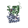
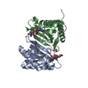
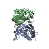
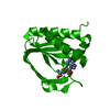
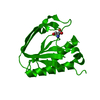
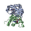
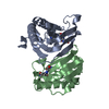
- PDB-2hl1: Crystal structure of the editing domain of threonyl-tRNA syntheta... -
+ Open data

- Basic information
| Entry | Database: PDB / ID: 2hl1 | ||||||
|---|---|---|---|---|---|---|---|
| Title | Crystal structure of the editing domain of threonyl-tRNA synthetase from Pyrococcus abyssi in complex with seryl-3'-aminoadenosine | ||||||
 Components Components | Threonyl-tRNA synthetase | ||||||
 Keywords Keywords | LIGASE / translation / editing / aminoacyl-tRNA synthetase / enzyme mechanism / enantioselectivity | ||||||
| Function / homology |  Function and homology information Function and homology informationthreonine-tRNA ligase / threonyl-tRNA aminoacylation / threonine-tRNA ligase activity / tRNA binding / zinc ion binding / ATP binding / cytosol Similarity search - Function | ||||||
| Biological species |   Pyrococcus abyssi (archaea) Pyrococcus abyssi (archaea) | ||||||
| Method |  X-RAY DIFFRACTION / MOLECULAR REPLACEMENT / Resolution: 2.25 Å X-RAY DIFFRACTION / MOLECULAR REPLACEMENT / Resolution: 2.25 Å | ||||||
 Authors Authors | Hussain, T. / Kruparani, S.P. / Pal, B. / Sankaranarayanan, R. | ||||||
 Citation Citation | Journal: Embo J. / Year: 2006 Title: Post-transfer editing mechanism of a D-aminoacyl-tRNA deacylase-like domain in threonyl-tRNA synthetase from archaea Authors: Hussain, T. / Kruparani, S.P. / Pal, B. / Dock-Bregeon, A.C. / Dwivedi, S. / Shekar, M.R. / Sureshbabu, K. / Sankaranarayanan, R. #1: Journal: Nat.Struct.Mol.Biol. / Year: 2005Title: A D-amino acid editing module coupled to the translational apparatus in archaea Authors: Dwivedi, S. / Kruparani, S.P. / Sankaranarayanan, R. | ||||||
| History |
|
- Structure visualization
| Structure viewer | Molecule: MolmilJmol/JSmol |
|---|
- Downloads & links
-Download
| PDBx/mmCIF format | 2hl1.cif.gz | 77.1 KB | Display | PDBx/mmCIF format |
|---|---|---|---|---|
| PDB format | pdb2hl1.ent.gz | 56.6 KB | Display | PDB format |
| PDBx/mmJSON format | 2hl1.json.gz | Tree view | PDBx/mmJSON format | |
| Others |  Other downloads Other downloads |
-Validation report
| Summary document | 2hl1_validation.pdf.gz | 985.7 KB | Display | wwPDB validaton report |
|---|---|---|---|---|
| Full document | 2hl1_full_validation.pdf.gz | 990.3 KB | Display | |
| Data in XML | 2hl1_validation.xml.gz | 16.1 KB | Display | |
| Data in CIF | 2hl1_validation.cif.gz | 22.6 KB | Display | |
| Arichive directory | https://data.pdbj.org/pub/pdb/validation_reports/hl/2hl1ftp://data.pdbj.org/pub/pdb/validation_reports/hl/2hl1 | HTTPS FTP |
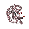
-Related structure data
| Related structure data |  2hkzC  2hl0C 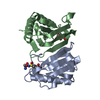 2hl2C  1y2qS S: Starting model for refinement C: citing same article ( |
|---|---|
| Similar structure data |
-Links
 PDBj
PDBj
- Assembly
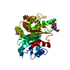
| Deposited unit | 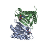
| ||||||||
|---|---|---|---|---|---|---|---|---|---|
| 1 |
| ||||||||
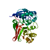
| Unit cell |
| ||||||||
| Details | The biological assembly is a dimer |
-Components
| #1: Protein | Mass: 16738.312 Da / Num. of mol.: 2 / Fragment: editing domain (residues 1-147) Source method: isolated from a genetically manipulated source Source: (gene. exp.) Pyrococcus abyssi (archaea) / Plasmid: pET21b / Species (production host): Escherichia coli / Production host:  #2: Chemical | 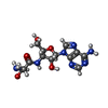  Mass: 353.334 Da / Num. of mol.: 2 / Source method: obtained synthetically / Formula: C13H19N7O5 Mass: 353.334 Da / Num. of mol.: 2 / Source method: obtained synthetically / Formula: C13H19N7O5#3: Water | ChemComp-HOH / |  Mass: 18.015 Da / Num. of mol.: 239 / Source method: isolated from a natural source / Formula: H2O Mass: 18.015 Da / Num. of mol.: 239 / Source method: isolated from a natural source / Formula: H2O |
|---|
-Experimental details
-Experiment
| Experiment | Method: X-RAY DIFFRACTION / Number of used crystals: 1 |
|---|
- Sample preparation
| Crystal | Density Matthews: 2.21 Å3/Da / Density % sol: 44.3 % |
|---|---|
| Crystal grow | Temperature: 277 K / Method: vapor diffusion, hanging drop / pH: 7 Details: 25% PEG 3350, 0.1M HEPES, pH 7.0, VAPOR DIFFUSION, HANGING DROP, temperature 277K |
-Data collection
| Diffraction | Mean temperature: 100 K |
|---|---|
| Diffraction source | Source: ROTATING ANODE / Type: RIGAKU RU300 / Wavelength: 1.5418 Å |
| Detector | Type: MAR scanner 345 mm plate / Detector: IMAGE PLATE / Date: Aug 5, 2005 / Details: Osmic mirrors |
| Radiation | Protocol: SINGLE WAVELENGTH / Monochromatic (M) / Laue (L): M / Scattering type: x-ray |
| Radiation wavelength | Wavelength: 1.5418 Å / Relative weight: 1 |
| Reflection | Resolution: 2.25→25 Å / Num. all: 13321 / Num. obs: 13321 / % possible obs: 90.7 % / Observed criterion σ(F): 0 / Observed criterion σ(I): 0 / Redundancy: 3.6 % / Biso Wilson estimate: 23.4 Å2 / Rmerge(I) obs: 0.061 / Net I/σ(I): 15.82 |
| Reflection shell | Resolution: 2.25→2.33 Å / Redundancy: 3.1 % / Rmerge(I) obs: 0.301 / Mean I/σ(I) obs: 2.7 / Num. unique all: 1201 / % possible all: 83.9 |
- Processing
| Software |
| ||||||||||||||||||||||||||||||||||||
|---|---|---|---|---|---|---|---|---|---|---|---|---|---|---|---|---|---|---|---|---|---|---|---|---|---|---|---|---|---|---|---|---|---|---|---|---|---|
| Refinement | Method to determine structure: MOLECULAR REPLACEMENT Starting model: PDB ENTRY 1Y2Q Resolution: 2.25→24.4 Å / Rfactor Rfree error: 0.011 / Data cutoff high absF: 1433959.68 / Data cutoff low absF: 0 / Isotropic thermal model: RESTRAINED / Cross valid method: THROUGHOUT / σ(F): 0 / Stereochemistry target values: Engh & Huber
| ||||||||||||||||||||||||||||||||||||
| Solvent computation | Solvent model: FLAT MODEL / Bsol: 40.7539 Å2 / ksol: 0.33344 e/Å3 | ||||||||||||||||||||||||||||||||||||
| Displacement parameters | Biso mean: 28.8 Å2
| ||||||||||||||||||||||||||||||||||||
| Refine analyze |
| ||||||||||||||||||||||||||||||||||||
| Refinement step | Cycle: LAST / Resolution: 2.25→24.4 Å
| ||||||||||||||||||||||||||||||||||||
| Refine LS restraints |
| ||||||||||||||||||||||||||||||||||||
| LS refinement shell | Resolution: 2.25→2.39 Å / Rfactor Rfree error: 0.031 / Total num. of bins used: 6
| ||||||||||||||||||||||||||||||||||||
| Xplor file |
|
