 Movie
Movie Controller

[English] 日本語
 Yorodumi
- PDB-2gu0: Crystal Structure of Human Rotavirus NSP2 (Group C / Bristol Strain) -
+ Open data

- Basic information
| Entry | Database: PDB / ID: 2gu0 | ||||||
|---|---|---|---|---|---|---|---|
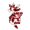
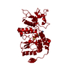
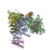
| Title | Crystal Structure of Human Rotavirus NSP2 (Group C / Bristol Strain) | ||||||
 Components Components | Nonstructural protein 2 | ||||||
 Keywords Keywords | VIRAL PROTEIN / NSP2 / rotavirus / HIT motif / Bristol | ||||||
| Function / homology |  Function and homology information Function and homology informationhydrolase activity, acting on acid anhydrides / viral genome replication / Hydrolases; Acting on acid anhydrides; Acting on acid anhydrides to facilitate cellular and subcellular movement / host cell cytoplasm / RNA binding / ATP binding / metal ion binding Similarity search - Function | ||||||
| Biological species |  Human rotavirus C Human rotavirus C | ||||||
| Method |  X-RAY DIFFRACTION / SYNCHROTRON / MOLECULAR REPLACEMENT / Resolution: 2.8 Å X-RAY DIFFRACTION / SYNCHROTRON / MOLECULAR REPLACEMENT / Resolution: 2.8 Å | ||||||
 Authors Authors | Jiang, X. / Prasad, B.V.V. | ||||||
 Citation Citation | Journal: J.Virol. / Year: 2006 Title: Structure-function analysis of rotavirus NSP2 octamer by using a novel complementation system Authors: Taraporewala, Z.F. / Jiang, X. / Vasquez-Del Carpio, R. / Jayaram, H. / Prasad, B.V.V. / Patton, J.T. | ||||||
| History |
|
- Structure visualization
| Structure viewer | Molecule: MolmilJmol/JSmol |
|---|
- Downloads & links
-Download
| PDBx/mmCIF format | 2gu0.cif.gz | 126.6 KB | Display | PDBx/mmCIF format |
|---|---|---|---|---|
| PDB format | pdb2gu0.ent.gz | 101.6 KB | Display | PDB format |
| PDBx/mmJSON format | 2gu0.json.gz | Tree view | PDBx/mmJSON format | |
| Others |  Other downloads Other downloads |
-Validation report
| Summary document | 2gu0_validation.pdf.gz | 421.1 KB | Display | wwPDB validaton report |
|---|---|---|---|---|
| Full document | 2gu0_full_validation.pdf.gz | 435.6 KB | Display | |
| Data in XML | 2gu0_validation.xml.gz | 23.2 KB | Display | |
| Data in CIF | 2gu0_validation.cif.gz | 31 KB | Display | |
| Arichive directory | https://data.pdbj.org/pub/pdb/validation_reports/gu/2gu0ftp://data.pdbj.org/pub/pdb/validation_reports/gu/2gu0 | HTTPS FTP |
-Related structure data
| Related structure data | |
|---|---|
| Similar structure data |


-Links
 PDBj
PDBj- Assembly
| Deposited unit | 
| ||||||||
|---|---|---|---|---|---|---|---|---|---|
| 1 | 
| ||||||||
| Unit cell |
| ||||||||
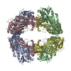
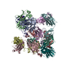
| Details | The biological assembly is a octamer generated from the two monomers in the asymmetric unit by the operations: (-x, -y, z), (-y, x, z) and (y, -x, z). |
-Components
| #1: Protein | Mass: 35964.145 Da / Num. of mol.: 2 Source method: isolated from a genetically manipulated source Source: (gene. exp.) Human rotavirus C / Species: Rotavirus C / Strain: Bristol / Gene: NSP2, segment 9 / Plasmid: pQE60 / Production host:  |
|---|
-Experimental details
-Experiment
| Experiment | Method: X-RAY DIFFRACTION / Number of used crystals: 1 |
|---|
- Sample preparation
| Crystal | Density Matthews: 3.51 Å3/Da / Density % sol: 64.98 % |
|---|---|
| Crystal grow | Temperature: 298 K / Method: vapor diffusion, hanging drop / pH: 6.4 Details: 1.5-1.7M ammonium sulfate, 0.1M MES, pH 6.4, VAPOR DIFFUSION, HANGING DROP, temperature 298K |
-Data collection
| Diffraction | Mean temperature: 100 K |
|---|---|
| Diffraction source | Source: SYNCHROTRON / Site: APS  / Beamline: 19-ID / Wavelength: 0.98 Å / Beamline: 19-ID / Wavelength: 0.98 Å |
| Detector | Type: ADSC QUANTUM 315 / Detector: CCD / Date: Jun 18, 2004 |
| Radiation | Monochromator: high-resolution double-crystal sagittal focusing Protocol: SINGLE WAVELENGTH / Monochromatic (M) / Laue (L): M / Scattering type: x-ray |
| Radiation wavelength | Wavelength: 0.98 Å / Relative weight: 1 |
| Reflection | Resolution: 2.8→500 Å / Num. all: 23492 / Num. obs: 23468 / % possible obs: 99.9 % / Observed criterion σ(I): 2 / Redundancy: 7.6 % / Rmerge(I) obs: 0.053 / Net I/σ(I): 30.5 |
| Reflection shell | Resolution: 2.8→2.9 Å / Redundancy: 7.6 % / Rmerge(I) obs: 0.286 / Mean I/σ(I) obs: 5.6 / % possible all: 100 |
- Processing
| Software |
| |||||||||||||||||||||||||
|---|---|---|---|---|---|---|---|---|---|---|---|---|---|---|---|---|---|---|---|---|---|---|---|---|---|---|
| Refinement | Method to determine structure: MOLECULAR REPLACEMENT / Resolution: 2.8→500 Å / σ(F): 0 / Stereochemistry target values: Engh & Huber
| |||||||||||||||||||||||||
| Solvent computation | Bsol: 19.957 Å2 | |||||||||||||||||||||||||
| Displacement parameters | Biso mean: 45.124 Å2
| |||||||||||||||||||||||||
| Refinement step | Cycle: LAST / Resolution: 2.8→500 Å
| |||||||||||||||||||||||||
| Refine LS restraints |
| |||||||||||||||||||||||||
| Xplor file |
|
