 Movie
Movie Controller

[English] 日本語
 Yorodumi
- PDB-2glm: Crystal structure of (3R)-Hydroxyacyl-Acyl Carrier Protein Dehydr... -
+ Open data

- Basic information
| Entry | Database: PDB / ID: 2glm | ||||||
|---|---|---|---|---|---|---|---|
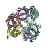
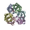
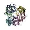
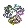
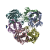
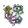
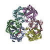
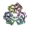
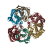
| Title | Crystal structure of (3R)-Hydroxyacyl-Acyl Carrier Protein Dehydratase(FabZ) from Helicobacter pylori complexed with Compound 2 | ||||||
 Components Components | (3R)-hydroxymyristoyl-acyl carrier protein dehydratase | ||||||
 Keywords Keywords | LYASE / FabZ complex | ||||||
| Function / homology |  Function and homology information Function and homology information3-hydroxyacyl-[acyl-carrier-protein] dehydratase / (3R)-hydroxyacyl-[acyl-carrier-protein] dehydratase activity / lipid A biosynthetic process / fatty acid biosynthetic process / membrane / cytoplasm Similarity search - Function | ||||||
| Biological species |   Helicobacter pylori (bacteria) Helicobacter pylori (bacteria) | ||||||
| Method |  X-RAY DIFFRACTION / MOLECULAR REPLACEMENT / Resolution: 2.6 Å X-RAY DIFFRACTION / MOLECULAR REPLACEMENT / Resolution: 2.6 Å | ||||||
 Authors Authors | Zhang, L. / Liu, W. / Shen, X. / Jiang, H. | ||||||
 Citation Citation | Journal: J.Biol.Chem. / Year: 2008 Title: Structural basis for catalytic and inhibitory mechanisms of beta-hydroxyacyl-acyl carrier protein dehydratase (FabZ). Authors: Zhang, L. / Liu, W. / Hu, T. / Du, L. / Luo, C. / Chen, K. / Shen, X. / Jiang, H. | ||||||
| History |
|
- Structure visualization
| Structure viewer | Molecule: MolmilJmol/JSmol |
|---|
- Downloads & links
-Download
| PDBx/mmCIF format | 2glm.cif.gz | 205.6 KB | Display | PDBx/mmCIF format |
|---|---|---|---|---|
| PDB format | pdb2glm.ent.gz | 163.9 KB | Display | PDB format |
| PDBx/mmJSON format | 2glm.json.gz | Tree view | PDBx/mmJSON format | |
| Others |  Other downloads Other downloads |
-Validation report
| Arichive directory | https://data.pdbj.org/pub/pdb/validation_reports/gl/2glmftp://data.pdbj.org/pub/pdb/validation_reports/gl/2glm | HTTPS FTP |
|---|
-Related structure data
| Related structure data |  2gllSC 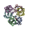 2glpC 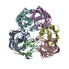 2glvC S: Starting model for refinement C: citing same article ( |
|---|---|
| Similar structure data |

-Links
 PDBj
PDBj

- Assembly
| Deposited unit | 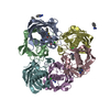
| ||||||||
|---|---|---|---|---|---|---|---|---|---|
| 1 |
| ||||||||
| Unit cell |
|
-Components
| #1: Protein | ( Mass: 19611.656 Da / Num. of mol.: 6 Source method: isolated from a genetically manipulated source Source: (gene. exp.) Helicobacter pylori (bacteria) / Strain: SS1 / Gene: FabZ / Plasmid: pQE30 / Production host: References: UniProt: Q5G940, UniProt: O25928*PLUS, Lyases; Carbon-oxygen lyases; Hydro-lyases #2: Chemical | ChemComp-CL /   Mass: 35.453 Da / Num. of mol.: 6 / Source method: obtained synthetically / Formula: Cl Mass: 35.453 Da / Num. of mol.: 6 / Source method: obtained synthetically / Formula: Cl#3: Chemical | ChemComp-BEN /   Mass: 120.152 Da / Num. of mol.: 7 / Source method: obtained synthetically / Formula: C7H8N2 Mass: 120.152 Da / Num. of mol.: 7 / Source method: obtained synthetically / Formula: C7H8N2#4: Chemical | ChemComp-SCB / |   Mass: 482.936 Da / Num. of mol.: 1 / Source method: obtained synthetically / Formula: C24H19ClN2O5S Mass: 482.936 Da / Num. of mol.: 1 / Source method: obtained synthetically / Formula: C24H19ClN2O5S#5: Water | ChemComp-HOH / |  Mass: 18.015 Da / Num. of mol.: 550 / Source method: isolated from a natural source / Formula: H2O Mass: 18.015 Da / Num. of mol.: 550 / Source method: isolated from a natural source / Formula: H2O |
|---|
-Experimental details
-Experiment
| Experiment | Method: X-RAY DIFFRACTION / Number of used crystals: 1 |
|---|
- Sample preparation
| Crystal | Density Matthews: 2.94 Å3/Da / Density % sol: 58.11 % |
|---|---|
| Crystal grow | Temperature: 277 K / Method: vapor diffusion, hanging drop / pH: 3.8 Details: 0.1M Sodium Acetate trihydrate PH3.8, 2.0M Sodium Formate, 20%(w/v) Benzamidine HCl, VAPOR DIFFUSION, HANGING DROP, temperature 277K |
-Data collection
| Diffraction | Mean temperature: 100 K |
|---|---|
| Diffraction source | Source: ROTATING ANODE / Type: RIGAKU / Wavelength: 1.5418 Å |
| Detector | Type: RIGAKU RAXIS IV / Detector: IMAGE PLATE / Date: Aug 15, 2005 |
| Radiation | Protocol: SINGLE WAVELENGTH / Monochromatic (M) / Laue (L): M / Scattering type: x-ray |
| Radiation wavelength | Wavelength: 1.5418 Å / Relative weight: 1 |
| Reflection | Resolution: 2.6→50 Å / Num. all: 45511 / Num. obs: 37248 / % possible obs: 85.7 % / Observed criterion σ(F): 0 / Observed criterion σ(I): 0 / Redundancy: 4.4 % / Rmerge(I) obs: 0.033 / Rsym value: 0.033 / Net I/σ(I): 34.66 |
| Reflection shell | Resolution: 2.6→2.68 Å / Redundancy: 4.3 % / Rmerge(I) obs: 0.077 / Mean I/σ(I) obs: 16.03 / Num. unique all: 4426 / Rsym value: 0.077 |
- Processing
| Software |
| ||||||||||||||||||||||||
|---|---|---|---|---|---|---|---|---|---|---|---|---|---|---|---|---|---|---|---|---|---|---|---|---|---|
| Refinement | Method to determine structure: MOLECULAR REPLACEMENT Starting model: 2GLL Resolution: 2.6→50 Å / σ(F): 0
| ||||||||||||||||||||||||
| Solvent computation | Bsol: 29.214 Å2 | ||||||||||||||||||||||||
| Displacement parameters | Biso mean: 16.002 Å2
| ||||||||||||||||||||||||
| Refinement step | Cycle: LAST / Resolution: 2.6→50 Å
| ||||||||||||||||||||||||
| Refine LS restraints |
| ||||||||||||||||||||||||
| Xplor file |
|
