 Movie
Movie Controller

[English] 日本語
 Yorodumi
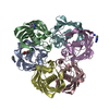
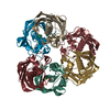
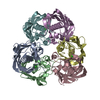
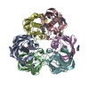
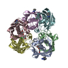
- PDB-3ed0: Crystal structure of (3R)-Hydroxyacyl-Acyl Carrier Protein Dehydr... -
+ Open data

- Basic information
| Entry | Database: PDB / ID: 3ed0 | ||||||
|---|---|---|---|---|---|---|---|
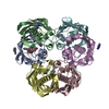
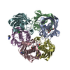
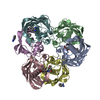
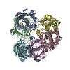
| Title | Crystal structure of (3R)-Hydroxyacyl-Acyl Carrier Protein Dehydratase (FabZ) from Helicobacter pylori in complex with emodin | ||||||
 Components Components | (3R)-hydroxymyristoyl-acyl carrier protein dehydratase | ||||||
 Keywords Keywords | LYASE / FabZ complex / Emodin / Cytoplasm / Lipid A biosynthesis / Lipid synthesis | ||||||
| Function / homology |  Function and homology information Function and homology information3-hydroxyacyl-[acyl-carrier-protein] dehydratase / (3R)-hydroxyacyl-[acyl-carrier-protein] dehydratase activity / lipid A biosynthetic process / fatty acid biosynthetic process / membrane / cytoplasm Similarity search - Function | ||||||
| Biological species |   Helicobacter pylori (bacteria) Helicobacter pylori (bacteria) | ||||||
| Method |  X-RAY DIFFRACTION / MOLECULAR REPLACEMENT / molecular replacement / Resolution: 2.3 Å X-RAY DIFFRACTION / MOLECULAR REPLACEMENT / molecular replacement / Resolution: 2.3 Å | ||||||
 Authors Authors | Zhang, L. / Zhang, H. / Liu, W. / Guo, Y. / Shen, X. / Jiang, H. | ||||||
 Citation Citation | Journal: BMC MICROBIOL. / Year: 2009 Title: Emodin targets the beta-hydroxyacyl-acyl carrier protein dehydratase from Helicobacter pylori: enzymatic inhibition assay with crystal structural and thermodynamic characterization Authors: Chen, J. / Zhang, L. / Zhang, Y. / Zhang, H. / Du, J. / Ding, J. / Guo, Y. / Jiang, H. / Shen, X. | ||||||
| History |
|
- Structure visualization
| Structure viewer | Molecule: MolmilJmol/JSmol |
|---|
- Downloads & links
-Download
| PDBx/mmCIF format | 3ed0.cif.gz | 200.1 KB | Display | PDBx/mmCIF format |
|---|---|---|---|---|
| PDB format | pdb3ed0.ent.gz | 159.6 KB | Display | PDB format |
| PDBx/mmJSON format | 3ed0.json.gz | Tree view | PDBx/mmJSON format | |
| Others |  Other downloads Other downloads |
-Validation report
| Arichive directory | https://data.pdbj.org/pub/pdb/validation_reports/ed/3ed0ftp://data.pdbj.org/pub/pdb/validation_reports/ed/3ed0 | HTTPS FTP |
|---|
-Related structure data
| Related structure data |  2gllS S: Starting model for refinement |
|---|---|
| Similar structure data |

-Links
 PDBj
PDBj

- Assembly
| Deposited unit | 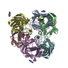
| ||||||||
|---|---|---|---|---|---|---|---|---|---|
| 1 |
| ||||||||
| Unit cell |
|
-Components
| #1: Protein | ( Mass: 18206.119 Da / Num. of mol.: 6 Source method: isolated from a genetically manipulated source Source: (gene. exp.) Helicobacter pylori (bacteria) / Strain: SS1 / Gene: fabZ / Plasmid: pQE30 / Production host: References: UniProt: Q5G940, Lyases; Carbon-oxygen lyases; Hydro-lyases #2: Chemical | ChemComp-BEN /   Mass: 120.152 Da / Num. of mol.: 7 / Source method: obtained synthetically / Formula: C7H8N2 Mass: 120.152 Da / Num. of mol.: 7 / Source method: obtained synthetically / Formula: C7H8N2#3: Chemical | ChemComp-CL /   Mass: 35.453 Da / Num. of mol.: 6 / Source method: obtained synthetically / Formula: Cl Mass: 35.453 Da / Num. of mol.: 6 / Source method: obtained synthetically / Formula: Cl#4: Chemical | 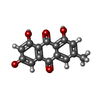  Mass: 270.237 Da / Num. of mol.: 2 / Source method: obtained synthetically / Formula: C15H10O5 Mass: 270.237 Da / Num. of mol.: 2 / Source method: obtained synthetically / Formula: C15H10O5#5: Water | ChemComp-HOH / |  Mass: 18.015 Da / Num. of mol.: 468 / Source method: isolated from a natural source / Formula: H2O Mass: 18.015 Da / Num. of mol.: 468 / Source method: isolated from a natural source / Formula: H2O |
|---|
-Experimental details
-Experiment
| Experiment | Method: X-RAY DIFFRACTION / Number of used crystals: 1 |
|---|
- Sample preparation
| Crystal | Density Matthews: 3.18 Å3/Da / Density % sol: 61.3 % |
|---|---|
| Crystal grow | Temperature: 277 K / Method: vapor diffusion, hanging drop / pH: 3.8 Details: 0.1M Sodium Acetate trihydrate, 2.0M Sodium Formate, 20%(w/v) Benzamidine HCl, pH 3.8, VAPOR DIFFUSION, HANGING DROP, temperature 277K |
-Data collection
| Diffraction | Mean temperature: 100 K | ||||||||||||||||||||||||||||||||||||||||||||||||||||||||||||||||||||||||||||||||||||||||
|---|---|---|---|---|---|---|---|---|---|---|---|---|---|---|---|---|---|---|---|---|---|---|---|---|---|---|---|---|---|---|---|---|---|---|---|---|---|---|---|---|---|---|---|---|---|---|---|---|---|---|---|---|---|---|---|---|---|---|---|---|---|---|---|---|---|---|---|---|---|---|---|---|---|---|---|---|---|---|---|---|---|---|---|---|---|---|---|---|---|
| Diffraction source | Source: ROTATING ANODE / Type: RIGAKU / Wavelength: 1.5418 Å | ||||||||||||||||||||||||||||||||||||||||||||||||||||||||||||||||||||||||||||||||||||||||
| Detector | Type: RIGAKU RAXIS IV / Detector: IMAGE PLATE / Date: Mar 15, 2008 | ||||||||||||||||||||||||||||||||||||||||||||||||||||||||||||||||||||||||||||||||||||||||
| Radiation | Protocol: SINGLE WAVELENGTH / Monochromatic (M) / Laue (L): M / Scattering type: x-ray | ||||||||||||||||||||||||||||||||||||||||||||||||||||||||||||||||||||||||||||||||||||||||
| Radiation wavelength | Wavelength: 1.5418 Å / Relative weight: 1 | ||||||||||||||||||||||||||||||||||||||||||||||||||||||||||||||||||||||||||||||||||||||||
| Reflection | Resolution: 2.3→93.25 Å / Num. obs: 62492 / % possible obs: 99.6 % / Redundancy: 8.9 % / Rmerge(I) obs: 0.124 / Rsym value: 0.124 / Net I/σ(I): 5.717 | ||||||||||||||||||||||||||||||||||||||||||||||||||||||||||||||||||||||||||||||||||||||||
| Reflection shell |
|
-Phasing
| Phasing | Method: molecular replacement |
|---|
- Processing
| Software |
| ||||||||||||||||||||||||||||||||||||
|---|---|---|---|---|---|---|---|---|---|---|---|---|---|---|---|---|---|---|---|---|---|---|---|---|---|---|---|---|---|---|---|---|---|---|---|---|---|
| Refinement | Method to determine structure: MOLECULAR REPLACEMENT Starting model: 2GLL Resolution: 2.3→20 Å / Occupancy max: 1 / Occupancy min: 0.8 / Cross valid method: THROUGHOUT
| ||||||||||||||||||||||||||||||||||||
| Displacement parameters | Biso max: 67.75 Å2 / Biso mean: 24.596 Å2 / Biso min: 9.25 Å2 | ||||||||||||||||||||||||||||||||||||
| Refinement step | Cycle: LAST / Resolution: 2.3→20 Å
|
