 Movie
Movie Controller

[English] 日本語
 Yorodumi
- PDB-2g7b: Crystal Structure of the R132K:R111L:L121E mutant of Cellular Ret... -
+ Open data

- Basic information
| Entry | Database: PDB / ID: 2g7b | ||||||
|---|---|---|---|---|---|---|---|
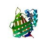
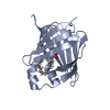
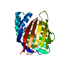
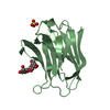
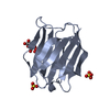
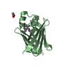
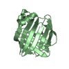
| Title | Crystal Structure of the R132K:R111L:L121E mutant of Cellular Retinoic Acid Binding Protein Type II In Complex With All-Trans-Retinal At 1.18 Angstroms Resolution | ||||||
 Components Components | Cellular retinoic acid-binding protein 2 | ||||||
 Keywords Keywords | TRANSPORT PROTEIN / CRABPII / Retinoic Acid / Retinoids / Beta Barrel / High Resolution / Schiff Base / Protonated Schiff Base / Retinal | ||||||
| Function / homology |  Function and homology information Function and homology informationpositive regulation of collateral sprouting / retinoid binding / retinal binding / retinoic acid binding / embryonic forelimb morphogenesis / retinoic acid metabolic process / retinol binding / Signaling by Retinoic Acid / cyclin binding / epidermis development ...positive regulation of collateral sprouting / retinoid binding / retinal binding / retinoic acid binding / embryonic forelimb morphogenesis / retinoic acid metabolic process / retinol binding / Signaling by Retinoic Acid / cyclin binding / epidermis development / fatty acid transport / fatty acid binding / regulation of DNA-templated transcription / signal transduction / endoplasmic reticulum / extracellular exosome / nucleoplasm / nucleus / cytoplasm / cytosol Similarity search - Function | ||||||
| Biological species |  Homo sapiens (human) Homo sapiens (human) | ||||||
| Method |  X-RAY DIFFRACTION / SYNCHROTRON / Rigid Body Refinement / Resolution: 1.18 Å X-RAY DIFFRACTION / SYNCHROTRON / Rigid Body Refinement / Resolution: 1.18 Å | ||||||
 Authors Authors | Vaezeslami, S. / Geiger, J.H. | ||||||
 Citation Citation | Journal: J.Am.Chem.Soc. / Year: 2007 Title: Protein design: reengineering cellular retinoic acid binding protein II into a rhodopsin protein mimic. Authors: Vasileiou, C. / Vaezeslami, S. / Crist, R.M. / Rabago-Smith, M. / Geiger, J.H. / Borhan, B. #1: Journal: ThesisTitle: determining crystal structures of proteins and protein complexes by X-ray crystallography: X-ray crystallographic studies of the mutants of cellular retinoic acid binding protein type II ...Title: determining crystal structures of proteins and protein complexes by X-ray crystallography: X-ray crystallographic studies of the mutants of cellular retinoic acid binding protein type II toward designing a mimic of rhodopsin Authors: Vaezeslami, S. | ||||||
| History |
| ||||||
| Remark 600 | Heterogen All-trans retinal (ligand code RET) formed a Protonated Schiff Base (PSB) with Lys 132 ...Heterogen All-trans retinal (ligand code RET) formed a Protonated Schiff Base (PSB) with Lys 132 and converted into all-trans axeropthene (ligand code AZE) |
- Structure visualization
| Structure viewer | Molecule: MolmilJmol/JSmol |
|---|
- Downloads & links
-Download
| PDBx/mmCIF format | 2g7b.cif.gz | 80.4 KB | Display | PDBx/mmCIF format |
|---|---|---|---|---|
| PDB format | pdb2g7b.ent.gz | 64.4 KB | Display | PDB format |
| PDBx/mmJSON format | 2g7b.json.gz | Tree view | PDBx/mmJSON format | |
| Others |  Other downloads Other downloads |
-Validation report
| Summary document | 2g7b_validation.pdf.gz | 630.6 KB | Display | wwPDB validaton report |
|---|---|---|---|---|
| Full document | 2g7b_full_validation.pdf.gz | 631.6 KB | Display | |
| Data in XML | 2g7b_validation.xml.gz | 11.2 KB | Display | |
| Data in CIF | 2g7b_validation.cif.gz | 17 KB | Display | |
| Arichive directory | https://data.pdbj.org/pub/pdb/validation_reports/g7/2g7bftp://data.pdbj.org/pub/pdb/validation_reports/g7/2g7b | HTTPS FTP |
-Related structure data
| Related structure data | 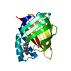 2g78C  2g79C  2g7a C: citing same article ( |
|---|---|
| Similar structure data |


-Links
 PDBj
PDBj


- Assembly
| Deposited unit | 
| ||||||||
|---|---|---|---|---|---|---|---|---|---|
| 1 |
| ||||||||
| Unit cell |
|
-Components
| #1: Protein | Mass: 15525.708 Da / Num. of mol.: 1 / Mutation: R132K, R111L, L121E Source method: isolated from a genetically manipulated source Source: (gene. exp.) Homo sapiens (human) / Gene: CRABP2 / Plasmid: pET17-b / Production host:  | ||||
|---|---|---|---|---|---|
| #2: Chemical |   Mass: 22.990 Da / Num. of mol.: 2 / Source method: obtained synthetically / Formula: Na Mass: 22.990 Da / Num. of mol.: 2 / Source method: obtained synthetically / Formula: Na#3: Chemical | ChemComp-AZE / |   Mass: 270.452 Da / Num. of mol.: 1 / Source method: obtained synthetically / Formula: C20H30 Mass: 270.452 Da / Num. of mol.: 1 / Source method: obtained synthetically / Formula: C20H30#4: Water | ChemComp-HOH / |  Mass: 18.015 Da / Num. of mol.: 250 / Source method: isolated from a natural source / Formula: H2O Mass: 18.015 Da / Num. of mol.: 250 / Source method: isolated from a natural source / Formula: H2O |
-Experimental details
-Experiment
| Experiment | Method: X-RAY DIFFRACTION / Number of used crystals: 1 |
|---|
- Sample preparation
| Crystal | Density Matthews: 2.59 Å3/Da / Density % sol: 52.5 % |
|---|---|
| Crystal grow | Temperature: 277 K / Method: vapor diffusion, hanging drop / pH: 5.8 Details: 18% (w/v) PEG 6000 and 0.1 M sodium acetate pH 5.8, VAPOR DIFFUSION, HANGING DROP, temperature 277K |
-Data collection
| Diffraction |
| |||||||||
|---|---|---|---|---|---|---|---|---|---|---|
| Diffraction source | Source: SYNCHROTRON / Site: APS  / Beamline: 32-ID / Wavelength: 1 Å / Beamline: 32-ID / Wavelength: 1 Å | |||||||||
| Detector | Type: MARRESEARCH / Detector: CCD / Date: Jun 19, 2004 | |||||||||
| Radiation | Protocol: SINGLE WAVELENGTH / Monochromatic (M) / Laue (L): M / Scattering type: x-ray | |||||||||
| Radiation wavelength | Wavelength: 1 Å / Relative weight: 1 | |||||||||
| Reflection | Resolution: 1.18→40 Å / Num. all: 54241 / Num. obs: 52457 / % possible obs: 96.7 % / Observed criterion σ(I): -3 / Rmerge(I) obs: 0.072 / Χ2: 0.567 / Net I/σ(I): 25.6 | |||||||||
| Reflection shell | Resolution: 1.18→1.22 Å / % possible obs: 74.6 % / Rmerge(I) obs: 0.332 / Num. unique obs: 4008 / Χ2: 0.395 / % possible all: 74.6 |
- Processing
| Software |
| |||||||||||||||||||||||||||||||||||||||||||||||||||||||||||||||||||||||||||||||||||||||||||||||||||||||||||||||||||||||||||||||||||||||||||||||||
|---|---|---|---|---|---|---|---|---|---|---|---|---|---|---|---|---|---|---|---|---|---|---|---|---|---|---|---|---|---|---|---|---|---|---|---|---|---|---|---|---|---|---|---|---|---|---|---|---|---|---|---|---|---|---|---|---|---|---|---|---|---|---|---|---|---|---|---|---|---|---|---|---|---|---|---|---|---|---|---|---|---|---|---|---|---|---|---|---|---|---|---|---|---|---|---|---|---|---|---|---|---|---|---|---|---|---|---|---|---|---|---|---|---|---|---|---|---|---|---|---|---|---|---|---|---|---|---|---|---|---|---|---|---|---|---|---|---|---|---|---|---|---|---|---|---|---|
| Refinement | Method to determine structure: Rigid Body Refinement / Resolution: 1.18→22.81 Å / Cor.coef. Fo:Fc: 0.979 / Cor.coef. Fo:Fc free: 0.972 / SU B: 0.899 / SU ML: 0.019 / Cross valid method: THROUGHOUT / σ(F): 0 / ESU R: 0.031 / ESU R Free: 0.032 / Stereochemistry target values: MAXIMUM LIKELIHOOD / Details: HYDROGENS HAVE BEEN ADDED IN THE RIDING POSITIONS
| |||||||||||||||||||||||||||||||||||||||||||||||||||||||||||||||||||||||||||||||||||||||||||||||||||||||||||||||||||||||||||||||||||||||||||||||||
| Solvent computation | Ion probe radii: 0.8 Å / Shrinkage radii: 0.8 Å / VDW probe radii: 1.2 Å / Solvent model: BABINET MODEL WITH MASK | |||||||||||||||||||||||||||||||||||||||||||||||||||||||||||||||||||||||||||||||||||||||||||||||||||||||||||||||||||||||||||||||||||||||||||||||||
| Displacement parameters | Biso mean: 12.18 Å2
| |||||||||||||||||||||||||||||||||||||||||||||||||||||||||||||||||||||||||||||||||||||||||||||||||||||||||||||||||||||||||||||||||||||||||||||||||
| Refinement step | Cycle: LAST / Resolution: 1.18→22.81 Å
| |||||||||||||||||||||||||||||||||||||||||||||||||||||||||||||||||||||||||||||||||||||||||||||||||||||||||||||||||||||||||||||||||||||||||||||||||
| Refine LS restraints |
| |||||||||||||||||||||||||||||||||||||||||||||||||||||||||||||||||||||||||||||||||||||||||||||||||||||||||||||||||||||||||||||||||||||||||||||||||
| LS refinement shell | Resolution: 1.18→1.208 Å / Total num. of bins used: 20
|
