 Movie
Movie Controller

[English] 日本語
 Yorodumi
- PDB-2g2d: Crystal structure of a putative pduO-type ATP:cobalamin adenosylt... -
+ Open data

- Basic information
| Entry | Database: PDB / ID: 2g2d | ||||||
|---|---|---|---|---|---|---|---|
| Title | Crystal structure of a putative pduO-type ATP:cobalamin adenosyltransferase from Mycobacterium tuberculosis | ||||||
 Components Components | ATP:cobalamin adenosyltransferase | ||||||
 Keywords Keywords | TRANSFERASE / helix bundle / trimer / Structural Genomics / PSI / Protein Structure Initiative / TB Structural Genomics Consortium / TBSGC | ||||||
| Function / homology |  Function and homology information Function and homology informationcorrinoid adenosyltransferase / corrinoid adenosyltransferase activity / porphyrin-containing compound biosynthetic process / cobalamin biosynthetic process / ATP binding / cytoplasm Similarity search - Function | ||||||
| Biological species |   Mycobacterium tuberculosis (bacteria) Mycobacterium tuberculosis (bacteria) | ||||||
| Method |  X-RAY DIFFRACTION / SYNCHROTRON / MAD / Resolution: 2 Å X-RAY DIFFRACTION / SYNCHROTRON / MAD / Resolution: 2 Å | ||||||
 Authors Authors | Moon, J.H. / Kaviratne, A. / Yu, M. / Bursey, E.H. / Hung, L.-W. / Lekin, T.P. / Segelke, B.W. / Terwilliger, T.C. / Kim, C.-Y. / TB Structural Genomics Consortium (TBSGC) | ||||||
 Citation Citation | Journal: To be Published Title: Crystal structure of a putative pduO-type ATP:cobalamin adenosyltransferase from Mycobacterium tuberculosis. Authors: Moon, J.H. / Kaviratne, A. / Yu, M. / Bursey, E.H. / Hung, L.-W. / Lekin, T.P. / Segelke, B.W. / Terwilliger, T.C. / Kim, C.-Y. | ||||||
| History |
|
- Structure visualization
| Structure viewer | Molecule: MolmilJmol/JSmol |
|---|
- Downloads & links
-Download
| PDBx/mmCIF format | 2g2d.cif.gz | 43.8 KB | Display | PDBx/mmCIF format |
|---|---|---|---|---|
| PDB format | pdb2g2d.ent.gz | 31.6 KB | Display | PDB format |
| PDBx/mmJSON format | 2g2d.json.gz | Tree view | PDBx/mmJSON format | |
| Others |  Other downloads Other downloads |
-Validation report
| Arichive directory | https://data.pdbj.org/pub/pdb/validation_reports/g2/2g2dftp://data.pdbj.org/pub/pdb/validation_reports/g2/2g2d | HTTPS FTP |
|---|
-Related structure data
| Similar structure data | |
|---|---|
| Other databases |


-Links
 PDBj
PDBj- Assembly
| Deposited unit | 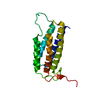
| ||||||||
|---|---|---|---|---|---|---|---|---|---|
| 1 | 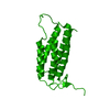
| ||||||||
| Unit cell |
| ||||||||
| Details | The biological assembly is a trimer generated by the operations: z, y, x and y, z, x. |
-Components
| #1: Protein | Mass: 20718.373 Da / Num. of mol.: 1 Source method: isolated from a genetically manipulated source Source: (gene. exp.) Mycobacterium tuberculosis (bacteria) / Plasmid: pET-28b / Species (production host): Escherichia coli / Production host: References: UniProt: P64803, UniProt: P9WP99*PLUS, corrinoid adenosyltransferase |
|---|---|
| #2: Water | ChemComp-HOH /  Mass: 18.015 Da / Num. of mol.: 79 / Source method: isolated from a natural source / Formula: H2O Mass: 18.015 Da / Num. of mol.: 79 / Source method: isolated from a natural source / Formula: H2O |
-Experimental details
-Experiment
| Experiment | Method: X-RAY DIFFRACTION / Number of used crystals: 1 |
|---|
- Sample preparation
| Crystal | Density Matthews: 2.28 Å3/Da / Density % sol: 45.95 % |
|---|---|
| Crystal grow | Temperature: 295 K / Method: vapor diffusion, hanging drop / pH: 6.5 Details: 0.1M MES, 1.6M Ammonium Sulfate, 10% Dioxane, pH 6.5, VAPOR DIFFUSION, HANGING DROP, temperature 295K |
-Data collection
| Diffraction |
| ||||||||||||||||||
|---|---|---|---|---|---|---|---|---|---|---|---|---|---|---|---|---|---|---|---|
| Diffraction source |
| ||||||||||||||||||
| Detector |
| ||||||||||||||||||
| Radiation |
| ||||||||||||||||||
| Radiation wavelength |
| ||||||||||||||||||
| Reflection | Resolution: 2→30 Å / Num. all: 13272 / Num. obs: 13238 / % possible obs: 99.2 % / Observed criterion σ(F): 0 / Observed criterion σ(I): 0 / Biso Wilson estimate: 24.1 Å2 | ||||||||||||||||||
| Reflection shell | Resolution: 2→2.07 Å / % possible all: 91.3 |

- Processing
| Software |
| ||||||||||||||||||||||||||||||||||||
|---|---|---|---|---|---|---|---|---|---|---|---|---|---|---|---|---|---|---|---|---|---|---|---|---|---|---|---|---|---|---|---|---|---|---|---|---|---|
| Refinement | Method to determine structure: MAD / Resolution: 2→19.8 Å / Rfactor Rfree error: 0.009 / Data cutoff high absF: 5552242.56 / Data cutoff low absF: 0 / Isotropic thermal model: RESTRAINED / Cross valid method: THROUGHOUT / σ(F): 0 / Stereochemistry target values: Engh & Huber
| ||||||||||||||||||||||||||||||||||||
| Solvent computation | Solvent model: FLAT MODEL / Bsol: 54.7764 Å2 / ksol: 0.356097 e/Å3 | ||||||||||||||||||||||||||||||||||||
| Displacement parameters | Biso mean: 33.1 Å2
| ||||||||||||||||||||||||||||||||||||
| Refine analyze |
| ||||||||||||||||||||||||||||||||||||
| Refinement step | Cycle: LAST / Resolution: 2→19.8 Å
| ||||||||||||||||||||||||||||||||||||
| Refine LS restraints |
| ||||||||||||||||||||||||||||||||||||
| LS refinement shell | Resolution: 2→2.13 Å / Rfactor Rfree error: 0.026 / Total num. of bins used: 6
| ||||||||||||||||||||||||||||||||||||
| Xplor file |
|
