 Movie
Movie Controller


+ Open data

- Basic information
| Entry | Database: PDB / ID: 1.0E+70 | ||||||||||||
|---|---|---|---|---|---|---|---|---|---|---|---|---|---|
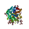
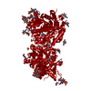
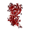
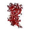
| Title | 2-F-glucosylated MYROSINASE FROM SINAPIS ALBA | ||||||||||||
 Components Components | MYROSINASE MA1 | ||||||||||||
 Keywords Keywords | HYDROLASE / FAMILY 1 GLYCOSYL HYDROLASE / GLUCOSINOLATE / MYROSINASE / TIM BARREL / GLUCOSYL ENZYME | ||||||||||||
| Function / homology |  Function and homology information Function and homology informationthioglucosidase / thioglucosidase activity / vacuole / beta-glucosidase activity / carbohydrate metabolic process / metal ion binding Similarity search - Function | ||||||||||||
| Biological species |  SINAPIS ALBA (white mustard) SINAPIS ALBA (white mustard) | ||||||||||||
| Method |  X-RAY DIFFRACTION / SYNCHROTRON / MOLECULAR REPLACEMENT / Resolution: 1.65 Å X-RAY DIFFRACTION / SYNCHROTRON / MOLECULAR REPLACEMENT / Resolution: 1.65 Å | ||||||||||||
 Authors Authors | Burmeister, W.P. | ||||||||||||
 Citation Citation | Journal: J.Biol.Chem. / Year: 2000 Title: High Resolution X-Ray Crystallography Shows that Ascorbate is a Cofactor for Myrosinase and Substitutes for the Function of the Catalytic Base Authors: Burmeister, W.P. / Cottaz, S. / Rollin, P. / Vasella, A. / Henrissat, B. #1: Journal: Structure / Year: 1997Title: The Crystal Structures of Sinapis Alba Myrosinase and a Covalent Glycosyl-Enzyme Intermediate Provide Insights Into the Substrate Recognition and Active-Site Machinery of an S-Glycosidase Authors: Burmeister, W.P. / Cottaz, S. / Driguez, H. / Iori, R. / Palmieri, S. / Henrissat, B. | ||||||||||||
| History |
|
- Structure visualization
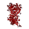
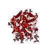
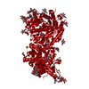
| Structure viewer | Molecule: MolmilJmol/JSmol |
|---|
- Downloads & links
-Download
| PDBx/mmCIF format | 1e70.cif.gz | 145.2 KB | Display | PDBx/mmCIF format |
|---|---|---|---|---|
| PDB format | pdb1e70.ent.gz | 113 KB | Display | PDB format |
| PDBx/mmJSON format | 1e70.json.gz | Tree view | PDBx/mmJSON format | |
| Others |  Other downloads Other downloads |
-Validation report
| Arichive directory | https://data.pdbj.org/pub/pdb/validation_reports/e7/1e70ftp://data.pdbj.org/pub/pdb/validation_reports/e7/1e70 | HTTPS FTP |
|---|
-Related structure data
| Related structure data |  1e4mSC  1e6qC  1e6sC  1e6xC  1e71C  1e72C  1e73C C: citing same article ( S: Starting model for refinement |
|---|---|
| Similar structure data |


-Links
 PDBj
PDBj- Assembly
| Deposited unit | 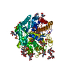
| |||||||||||||||
|---|---|---|---|---|---|---|---|---|---|---|---|---|---|---|---|---|
| 1 | 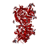
| |||||||||||||||
| Unit cell |
| |||||||||||||||
| Components on special symmetry positions |
|
-Components
-Protein , 1 types, 1 molecules M
| #1: Protein | Mass: 57078.289 Da / Num. of mol.: 1 / Source method: isolated from a natural source / Source: (natural) SINAPIS ALBA (white mustard) / Cellular location: MYROSIN GRAINS / Organ: SEED / Strain: EMERGO / References: UniProt: P29736, thioglucosidase |
|---|
-Sugars , 5 types, 10 molecules 

| #2: Polysaccharide | 2-acetamido-2-deoxy-beta-D-glucopyranose-(1-4)-2-acetamido-2-deoxy-beta-D-glucopyranose Source method: isolated from a genetically manipulated source | ||
|---|---|---|---|
| #3: Polysaccharide | beta-D-xylopyranose-(1-2)-beta-D-mannopyranose-(1-4)-2-acetamido-2-deoxy-beta-D-glucopyranose-(1-4)- ...beta-D-xylopyranose-(1-2)-beta-D-mannopyranose-(1-4)-2-acetamido-2-deoxy-beta-D-glucopyranose-(1-4)-[alpha-L-fucopyranose-(1-3)]2-acetamido-2-deoxy-beta-D-glucopyranose Source method: isolated from a genetically manipulated source | ||
| #4: Polysaccharide | beta-D-xylopyranose-(1-2)-[alpha-D-mannopyranose-(1-3)]alpha-D-mannopyranose-(1-4)-2-acetamido-2- ...beta-D-xylopyranose-(1-2)-[alpha-D-mannopyranose-(1-3)]alpha-D-mannopyranose-(1-4)-2-acetamido-2-deoxy-beta-D-glucopyranose-(1-4)-[alpha-L-fucopyranose-(1-3)]2-acetamido-2-deoxy-beta-D-glucopyranose Source method: isolated from a genetically manipulated source | ||
| #5: Sugar | ChemComp-NAG /  Type: D-saccharide, beta linking / Mass: 221.208 Da / Num. of mol.: 6 Type: D-saccharide, beta linking / Mass: 221.208 Da / Num. of mol.: 6Source method: isolated from a genetically manipulated source Formula: C8H15NO6 #6: Sugar | ChemComp-G2F / |  Type: D-saccharide, alpha linking / Mass: 182.147 Da / Num. of mol.: 1 Type: D-saccharide, alpha linking / Mass: 182.147 Da / Num. of mol.: 1Source method: isolated from a genetically manipulated source Formula: C6H11FO5 |
-Non-polymers , 4 types, 807 molecules 


| #7: Chemical | ChemComp-ZN /  Mass: 65.409 Da / Num. of mol.: 1 / Source method: obtained synthetically / Formula: Zn Mass: 65.409 Da / Num. of mol.: 1 / Source method: obtained synthetically / Formula: Zn | ||||
|---|---|---|---|---|---|
| #8: Chemical | ChemComp-SO4 /  Mass: 96.063 Da / Num. of mol.: 8 / Source method: obtained synthetically / Formula: SO4 Mass: 96.063 Da / Num. of mol.: 8 / Source method: obtained synthetically / Formula: SO4#9: Chemical | ChemComp-GOL /  Mass: 92.094 Da / Num. of mol.: 4 / Source method: obtained synthetically / Formula: C3H8O3 Mass: 92.094 Da / Num. of mol.: 4 / Source method: obtained synthetically / Formula: C3H8O3#10: Water | ChemComp-HOH / | Mass: 18.015 Da / Num. of mol.: 794 / Source method: isolated from a natural source / Formula: H2O |
-Details
| Compound details | ACTIVE SITE NUCLEOPHIL| Has protein modification | Y | |
|---|
-Experimental details
-Experiment
| Experiment | Method: X-RAY DIFFRACTION / Number of used crystals: 1 |
|---|
- Sample preparation
| Crystal | Density Matthews: 3.2 Å3/Da / Density % sol: 50 % / Description: ONLY THE LIGAND DIFFERS | ||||||||||||||||||||||||||||||||||||
|---|---|---|---|---|---|---|---|---|---|---|---|---|---|---|---|---|---|---|---|---|---|---|---|---|---|---|---|---|---|---|---|---|---|---|---|---|---|
| Crystal grow | Method: vapor diffusion, hanging drop / pH: 7 Details: HANGING DROP METHOD, 12 MG/ML PROTEIN IN 30 MM HEPES, PH 6.5, 0.05 % NAN3 PRECIPITANT 66 % SAT. AMMONIUM SULFATE, 100MM TRIS-HCL PH 7.0 | ||||||||||||||||||||||||||||||||||||
| Crystal grow | *PLUS pH: 6.5 / Method: vapor diffusion, hanging dropDetails: Burmeister, W.P., (1997) Structure (London), 5, 663. | ||||||||||||||||||||||||||||||||||||
| Components of the solutions | *PLUS
|
-Data collection
| Diffraction | Mean temperature: 100 K |
|---|---|
| Diffraction source | Source: SYNCHROTRON / Site: ESRF  / Beamline: ID14-3 / Wavelength: 0.935 / Beamline: ID14-3 / Wavelength: 0.935 |
| Detector | Type: MARRESEARCH / Detector: CCD / Date: Nov 15, 1997 / Details: BENT MULTILAYER, SAGITALLY FOCUSING CRYSTAL |
| Radiation | Monochromator: DIAMOND C(111) / Protocol: SINGLE WAVELENGTH / Monochromatic (M) / Laue (L): M / Scattering type: x-ray |
| Radiation wavelength | Wavelength: 0.935 Å / Relative weight: 1 |
| Reflection | Resolution: 1.65→34.7 Å / Num. obs: 90017 / % possible obs: 88.5 % / Observed criterion σ(I): 0 / Redundancy: 3.3 % / Rmerge(I) obs: 0.085 / Rsym value: 0.085 / Net I/σ(I): 5 |
| Reflection shell | Resolution: 1.65→1.74 Å / Redundancy: 3 % / Rmerge(I) obs: 0.35 / Mean I/σ(I) obs: 1.8 / Rsym value: 0.35 / % possible all: 76.2 |
| Reflection shell | *PLUS % possible obs: 76.2 % / Redundancy: 2.4 % / Rmerge(I) obs: 0.35 |
- Processing
| Software |
| ||||||||||||||||||||||||||||||||||||||||||||||||||||||||||||||||||||||||||||||||||||
|---|---|---|---|---|---|---|---|---|---|---|---|---|---|---|---|---|---|---|---|---|---|---|---|---|---|---|---|---|---|---|---|---|---|---|---|---|---|---|---|---|---|---|---|---|---|---|---|---|---|---|---|---|---|---|---|---|---|---|---|---|---|---|---|---|---|---|---|---|---|---|---|---|---|---|---|---|---|---|---|---|---|---|---|---|---|
| Refinement | Method to determine structure: MOLECULAR REPLACEMENT Starting model: PDB ENTRY 1E4M Resolution: 1.65→10 Å / SU B: 1.8 / SU ML: 0.058 / Cross valid method: THROUGHOUT / σ(F): 0 / ESU R: 0.099 / ESU R Free: 0.096
| ||||||||||||||||||||||||||||||||||||||||||||||||||||||||||||||||||||||||||||||||||||
| Displacement parameters | Biso mean: 31.8 Å2 | ||||||||||||||||||||||||||||||||||||||||||||||||||||||||||||||||||||||||||||||||||||
| Refinement step | Cycle: LAST / Resolution: 1.65→10 Å
| ||||||||||||||||||||||||||||||||||||||||||||||||||||||||||||||||||||||||||||||||||||
| Refine LS restraints |
| ||||||||||||||||||||||||||||||||||||||||||||||||||||||||||||||||||||||||||||||||||||
| Software | *PLUS Name: REFMAC / Classification: refinement | ||||||||||||||||||||||||||||||||||||||||||||||||||||||||||||||||||||||||||||||||||||
| Refinement | *PLUS Rfactor obs: 0.169 | ||||||||||||||||||||||||||||||||||||||||||||||||||||||||||||||||||||||||||||||||||||
| Solvent computation | *PLUS | ||||||||||||||||||||||||||||||||||||||||||||||||||||||||||||||||||||||||||||||||||||
| Displacement parameters | *PLUS | ||||||||||||||||||||||||||||||||||||||||||||||||||||||||||||||||||||||||||||||||||||
| LS refinement shell | *PLUS Highest resolution: 1.65 Å / Lowest resolution: 1.74 Å / Rfactor Rfree: 0.34 / Rfactor obs: 0.293 |
