 Movie
Movie Controller

[English] 日本語
 Yorodumi
- PDB-1w9d: S. alba myrosinase in complex with S-ethyl phenylacetothiohydroxi... -
+ Open data

- Basic information
| Entry | Database: PDB / ID: 1w9d | |||||||||
|---|---|---|---|---|---|---|---|---|---|---|
| Title | S. alba myrosinase in complex with S-ethyl phenylacetothiohydroximate- O-sulfate | |||||||||
 Components Components | GLYCOSIDASE | |||||||||
 Keywords Keywords | HYDROLASE / THIOGLUCOSIDASE / THIOGLYCOSIDASE / MYROSINASE / THIO-GLUCOSIDE / THIOHYDROXIMATE / GLUSOSINOLATE / GLUCOTROPAEOLIN | |||||||||
| Function / homology |  Function and homology information Function and homology informationthioglucosidase / thioglucosidase activity / vacuole / beta-glucosidase activity / carbohydrate metabolic process / metal ion binding Similarity search - Function | |||||||||
| Biological species |  SINAPIS ALBA (white mustard) SINAPIS ALBA (white mustard) | |||||||||
| Method |  X-RAY DIFFRACTION / SYNCHROTRON / MOLECULAR REPLACEMENT / Resolution: 1.6 Å X-RAY DIFFRACTION / SYNCHROTRON / MOLECULAR REPLACEMENT / Resolution: 1.6 Å | |||||||||
 Authors Authors | Bourderioux, A. / Lefoix, M. / Gueyrard, D. / Tatibouet, A. / Cottaz, S. / Arzt, S. / Burmeister, W.P. / Rollin, P. | |||||||||
 Citation Citation | Journal: Org.Biomol.Chem. / Year: 2005 Title: The Glucosinolate-Myrosinase System. New Insights Into Enzyme-Substrate Interactions by Use of Simplified Inhibitors Authors: Bourderioux, A. / Lefoix, M. / Gueyrard, D. / Tatibouet, A. / Cottaz, S. / Arzt, S. / Burmeister, W.P. / Rollin, P. #1: Journal: J.Biol.Chem. / Year: 2000Title: High Resolution X-Ray Crystallography Shows that Ascorbate is a Cofactor for Myrosinase and Substitutes for the Function of the Catalytic Base Authors: Burmeister, W.P. / Cottaz, S. / Rollin, P. / Vasella, A. / Henrissat, B. | |||||||||
| History |
| |||||||||
| Remark 700 | SHEET THE SHEET STRUCTURE OF THIS MOLECULE IS BIFURCATED. IN ORDER TO REPRESENT THIS FEATURE IN ... SHEET THE SHEET STRUCTURE OF THIS MOLECULE IS BIFURCATED. IN ORDER TO REPRESENT THIS FEATURE IN THE SHEET RECORDS BELOW, TWO SHEETS ARE DEFINED. |
- Structure visualization
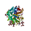
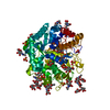
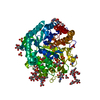
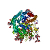
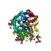
| Structure viewer | Molecule: MolmilJmol/JSmol |
|---|
- Downloads & links
-Download
| PDBx/mmCIF format | 1w9d.cif.gz | 142.3 KB | Display | PDBx/mmCIF format |
|---|---|---|---|---|
| PDB format | pdb1w9d.ent.gz | 110.7 KB | Display | PDB format |
| PDBx/mmJSON format | 1w9d.json.gz | Tree view | PDBx/mmJSON format | |
| Others |  Other downloads Other downloads |
-Validation report
| Arichive directory | https://data.pdbj.org/pub/pdb/validation_reports/w9/1w9dftp://data.pdbj.org/pub/pdb/validation_reports/w9/1w9d | HTTPS FTP |
|---|
-Related structure data
| Related structure data | 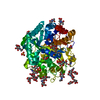 1w9bC  1e4mS C: citing same article ( S: Starting model for refinement |
|---|---|
| Similar structure data |


-Links
 PDBj
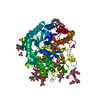
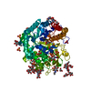
PDBj- Assembly
| Deposited unit | 
| |||||||||||||||
|---|---|---|---|---|---|---|---|---|---|---|---|---|---|---|---|---|
| 1 |
| |||||||||||||||
| Unit cell |
| |||||||||||||||
| Components on special symmetry positions |
|
-Components
-Protein , 1 types, 1 molecules M
| #1: Protein | Mass: 57078.289 Da / Num. of mol.: 1 / Source method: isolated from a natural source / Source: (natural) SINAPIS ALBA (white mustard) / Organ: SEED / Strain: EMERGO / Tissue: MYROSIN GRAINS / References: UniProt: P29736*PLUS, EC: 3.2.3.1 |
|---|
-Sugars , 4 types, 10 molecules 
| #2: Polysaccharide | Source method: isolated from a genetically manipulated source #3: Polysaccharide | beta-D-xylopyranose-(1-2)-beta-D-mannopyranose-(1-4)-2-acetamido-2-deoxy-beta-D-glucopyranose-(1-4)- ...beta-D-xylopyranose-(1-2)-beta-D-mannopyranose-(1-4)-2-acetamido-2-deoxy-beta-D-glucopyranose-(1-4)-[alpha-L-fucopyranose-(1-3)]2-acetamido-2-deoxy-beta-D-glucopyranose | Source method: isolated from a genetically manipulated source #4: Polysaccharide | beta-D-xylopyranose-(1-2)-[alpha-D-mannopyranose-(1-3)][alpha-D-mannopyranose-(1-6)]beta-D- ...beta-D-xylopyranose-(1-2)-[alpha-D-mannopyranose-(1-3)][alpha-D-mannopyranose-(1-6)]beta-D-mannopyranose-(1-4)-2-acetamido-2-deoxy-beta-D-glucopyranose-(1-4)-[alpha-L-fucopyranose-(1-3)]2-acetamido-2-deoxy-beta-D-glucopyranose | Source method: isolated from a genetically manipulated source #5: Sugar | ChemComp-NAG /  Type: D-saccharide, beta linking / Mass: 221.208 Da / Num. of mol.: 6 Type: D-saccharide, beta linking / Mass: 221.208 Da / Num. of mol.: 6Source method: isolated from a genetically manipulated source Formula: C8H15NO6 |
|---|
-Non-polymers , 5 types, 770 molecules 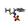


| #6: Chemical | ChemComp-SEH /  Mass: 275.345 Da / Num. of mol.: 1 / Source method: obtained synthetically / Formula: C10H13NO4S2 Mass: 275.345 Da / Num. of mol.: 1 / Source method: obtained synthetically / Formula: C10H13NO4S2 | ||||
|---|---|---|---|---|---|
| #7: Chemical | ChemComp-ZN /  Mass: 65.409 Da / Num. of mol.: 1 / Source method: obtained synthetically / Formula: Zn Mass: 65.409 Da / Num. of mol.: 1 / Source method: obtained synthetically / Formula: Zn | ||||
| #8: Chemical | ChemComp-SO4 /  Mass: 96.063 Da / Num. of mol.: 4 / Source method: obtained synthetically / Formula: SO4 Mass: 96.063 Da / Num. of mol.: 4 / Source method: obtained synthetically / Formula: SO4#9: Chemical |  Mass: 92.094 Da / Num. of mol.: 3 / Source method: obtained synthetically / Formula: C3H8O3 Mass: 92.094 Da / Num. of mol.: 3 / Source method: obtained synthetically / Formula: C3H8O3#10: Water | ChemComp-HOH / | Mass: 18.015 Da / Num. of mol.: 761 / Source method: isolated from a natural source / Formula: H2O |
-Details
| Has protein modification | Y |
|---|---|
| Sequence details | OUR SEQUENCE IS AN X-RAY SEQUENCE BASED ON THE ASSIGNMENT OF THE RESIDUES BY THEIR ELECTRON DENSITY. ...OUR SEQUENCE IS AN X-RAY SEQUENCE BASED ON THE ASSIGNMENT |
-Experimental details
-Experiment
| Experiment | Method: X-RAY DIFFRACTION / Number of used crystals: 1 |
|---|
- Sample preparation
| Crystal | Density Matthews: 3.2 Å3/Da / Density % sol: 50 % |
|---|---|
| Crystal grow | Method: vapor diffusion, hanging drop / pH: 7 Details: HANGING DROP METHOD, 12 MG/ML PROTEIN IN 30 MM HEPES, PH 6.5, 0.05 % NAN3, PRECIPITANT 66% SAT. AMMONIUM SULFATE, 100MM TRIS-HCL PH 7.0 |
-Data collection
| Diffraction | Mean temperature: 100 K |
|---|---|
| Diffraction source | Source: SYNCHROTRON / Site: ESRF  / Beamline: ID14-2 / Wavelength: 0.931 / Beamline: ID14-2 / Wavelength: 0.931 |
| Detector | Type: MARRESEARCH / Detector: CCD / Date: Sep 27, 2002 / Details: TOROIDAL MIRROR |
| Radiation | Monochromator: DIAMOND 111 AND SI220 / Protocol: SINGLE WAVELENGTH / Monochromatic (M) / Laue (L): M / Scattering type: x-ray |
| Radiation wavelength | Wavelength: 0.931 Å / Relative weight: 1 |
| Reflection | Resolution: 1.4→35 Å / Num. obs: 146755 / % possible obs: 99 % / Observed criterion σ(I): 0 / Redundancy: 4.4 % / Biso Wilson estimate: 12.6 Å2 / Rmerge(I) obs: 0.05 / Net I/σ(I): 6.1 |
| Reflection shell | Resolution: 1.4→1.48 Å / Redundancy: 3.9 % / Rmerge(I) obs: 0.32 / Mean I/σ(I) obs: 1.4 / % possible all: 97.4 |
- Processing
| Software |
| ||||||||||||||||||||||||||||||||||||||||||||||||||||||||||||||||||||||||||||||||
|---|---|---|---|---|---|---|---|---|---|---|---|---|---|---|---|---|---|---|---|---|---|---|---|---|---|---|---|---|---|---|---|---|---|---|---|---|---|---|---|---|---|---|---|---|---|---|---|---|---|---|---|---|---|---|---|---|---|---|---|---|---|---|---|---|---|---|---|---|---|---|---|---|---|---|---|---|---|---|---|---|---|
| Refinement | Method to determine structure: MOLECULAR REPLACEMENT Starting model: PDB ENTRY 1E4M Resolution: 1.6→35 Å / Rfactor Rfree error: 0.002 / Data cutoff high absF: 10000 / Isotropic thermal model: RESTRAINED / Cross valid method: THROUGHOUT / σ(F): 0 / Stereochemistry target values: MAXIMUM LIKELIHOOD Details: INHIBITOR TOPOLOGY AND PARAMETERS SAME AS IN ENTRY 1W9B, ONLY MOST OF THE ATOMS OF THE GLUCOSE GROUP HAVE BEEN DELETED. THE INHIBITOR HAS BEEN RENAMED FROM CGT TO SEH AFTER REFINEMENT
| ||||||||||||||||||||||||||||||||||||||||||||||||||||||||||||||||||||||||||||||||
| Solvent computation | Solvent model: FLAT MODEL / Bsol: 36.9271 Å2 / ksol: 0.380436 e/Å3 | ||||||||||||||||||||||||||||||||||||||||||||||||||||||||||||||||||||||||||||||||
| Displacement parameters | Biso mean: 18.46 Å2
| ||||||||||||||||||||||||||||||||||||||||||||||||||||||||||||||||||||||||||||||||
| Refine analyze |
| ||||||||||||||||||||||||||||||||||||||||||||||||||||||||||||||||||||||||||||||||
| Refinement step | Cycle: LAST / Resolution: 1.6→35 Å
| ||||||||||||||||||||||||||||||||||||||||||||||||||||||||||||||||||||||||||||||||
| Refine LS restraints |
| ||||||||||||||||||||||||||||||||||||||||||||||||||||||||||||||||||||||||||||||||
| LS refinement shell | Resolution: 1.6→1.66 Å / Rfactor Rfree error: 0.01 / Total num. of bins used: 10
| ||||||||||||||||||||||||||||||||||||||||||||||||||||||||||||||||||||||||||||||||
| Xplor file |
|
