 Movie
Movie Controller

[English] 日本語
 Yorodumi
- PDB-2f91: 1.2A resolution structure of a crayfish trypsin complexed with a ... -
+ Open data

- Basic information
| Entry | Database: PDB / ID: 2f91 | |||||||||
|---|---|---|---|---|---|---|---|---|---|---|
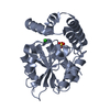
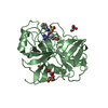
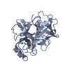
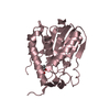
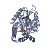
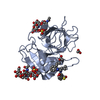
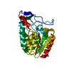
| Title | 1.2A resolution structure of a crayfish trypsin complexed with a peptide inhibitor, SGTI | |||||||||
 Components Components |
| |||||||||
 Keywords Keywords | HYDROLASE/HYDROLASE INHIBITOR / SERINE PROTEASE / TRYPSIN / CANONICAL INHIBITOR / ATOMIC RESOLUTION / HYDROLASE-HYDROLASE INHIBITOR COMPLEX | |||||||||
| Function / homology |  Function and homology information Function and homology informationserine-type endopeptidase inhibitor activity / serine-type endopeptidase activity / proteolysis / extracellular region / metal ion binding Similarity search - Function | |||||||||
| Biological species |  Pontastacus leptodactylus (narrow-clawed crayfish) Pontastacus leptodactylus (narrow-clawed crayfish) | |||||||||
| Method |  X-RAY DIFFRACTION / SYNCHROTRON / MOLECULAR REPLACEMENT / Resolution: 1.2 Å X-RAY DIFFRACTION / SYNCHROTRON / MOLECULAR REPLACEMENT / Resolution: 1.2 Å | |||||||||
 Authors Authors | Fodor, K. / Harmat, V. / Hetenyi, C. / Kardos, J. / Antal, J. / Perczel, A. / Patthy, A. / Katona, G. / Graf, L. | |||||||||
 Citation Citation | Journal: Biochemistry / Year: 2006 Title: Enzyme:Substrate Hydrogen Bond Shortening during the Acylation Phase of Serine Protease Catalysis. Authors: Fodor, K. / Harmat, V. / Neutze, R. / Szilagyi, L. / Graf, L. / Katona, G. #1: Journal: J.Mol.Biol. / Year: 2005 Title: Extended Intermolecular Interactions in a Serine Protease-Canonical Inhibitor Complex Account for Strong and Highly Specific Inhibition. Authors: Fodor, K. / Harmat, V. / Hetenyi, C. / Kardos, J. / Antal, J. / Perczel, A. / Patthy, A. / Katona, G. / Graf, L. | |||||||||
| History |
|
- Structure visualization
| Structure viewer | Molecule: MolmilJmol/JSmol |
|---|
- Downloads & links
-Download
| PDBx/mmCIF format | 2f91.cif.gz | 169.7 KB | Display | PDBx/mmCIF format |
|---|---|---|---|---|
| PDB format | pdb2f91.ent.gz | 135 KB | Display | PDB format |
| PDBx/mmJSON format | 2f91.json.gz | Tree view | PDBx/mmJSON format | |
| Others |  Other downloads Other downloads |
-Validation report
| Summary document | 2f91_validation.pdf.gz | 436.8 KB | Display | wwPDB validaton report |
|---|---|---|---|---|
| Full document | 2f91_full_validation.pdf.gz | 439.8 KB | Display | |
| Data in XML | 2f91_validation.xml.gz | 16.2 KB | Display | |
| Data in CIF | 2f91_validation.cif.gz | 24.3 KB | Display | |
| Arichive directory | https://data.pdbj.org/pub/pdb/validation_reports/f9/2f91ftp://data.pdbj.org/pub/pdb/validation_reports/f9/2f91 | HTTPS FTP |
-Related structure data
| Related structure data | 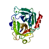 1h4wS S: Starting model for refinement |
|---|---|
| Similar structure data |


-Links
 PDBj
PDBj
- Assembly
| Deposited unit | 
| ||||||||
|---|---|---|---|---|---|---|---|---|---|
| 1 |
| ||||||||
| Unit cell |
|
-Components
| #1: Protein | Mass: 25072.434 Da / Num. of mol.: 1 / Source method: isolated from a natural source Source: (natural) Pontastacus leptodactylus (narrow-clawed crayfish)Tissue: HEPATOPANCREAS / References: UniProt: Q52V24, trypsin | ||||
|---|---|---|---|---|---|
| #2: Protein/peptide | Mass: 3828.314 Da / Num. of mol.: 1 / Fragment: PROTEASE INHIBITOR SGPI-1, RESIDUES 20-54 / Source method: obtained synthetically Details: THE PROTEIN WAS CHEMICALLY SYNTHESIZED, THIS SEQUENCE OCCURS NATURALLY IN SCHISTOCERCA GREGARIA (DESERT LOCUST) References: UniProt: O46162 | ||||
| #3: Chemical | ChemComp-CD /   Mass: 112.411 Da / Num. of mol.: 6 / Source method: obtained synthetically / Formula: Cd Mass: 112.411 Da / Num. of mol.: 6 / Source method: obtained synthetically / Formula: Cd#4: Chemical |   Mass: 35.453 Da / Num. of mol.: 2 / Source method: obtained synthetically / Formula: Cl Mass: 35.453 Da / Num. of mol.: 2 / Source method: obtained synthetically / Formula: Cl#5: Water | ChemComp-HOH / |  Mass: 18.015 Da / Num. of mol.: 343 / Source method: isolated from a natural source / Formula: H2O Mass: 18.015 Da / Num. of mol.: 343 / Source method: isolated from a natural source / Formula: H2O |
-Experimental details
-Experiment
| Experiment | Method: X-RAY DIFFRACTION / Number of used crystals: 1 |
|---|
- Sample preparation
| Crystal | Density Matthews: 2.07 Å3/Da / Density % sol: 40.65 % |
|---|---|
| Crystal grow | Temperature: 293 K / Method: vapor diffusion, hanging drop / pH: 4.6 Details: 30% PEG 400, 0.1 M CD CHLORIDE, 0.1 M NA ACETATE, pH 4.60, VAPOR DIFFUSION, HANGING DROP, temperature 293K |
-Data collection
| Diffraction | Mean temperature: 100 K |
|---|---|
| Diffraction source | Source: SYNCHROTRON / Site: ESRF  / Beamline: ID14-2 / Wavelength: 0.933 / Wavelength: 0.933 Å / Beamline: ID14-2 / Wavelength: 0.933 / Wavelength: 0.933 Å |
| Detector | Type: ADSC QUANTUM 4 / Detector: CCD / Details: MIRROR |
| Radiation | Monochromator: DIAMOND (111), GE (220) / Protocol: SINGLE WAVELENGTH / Monochromatic (M) / Laue (L): M / Scattering type: x-ray |
| Radiation wavelength | Wavelength: 0.933 Å / Relative weight: 1 |
| Reflection | Resolution: 1.2→32.1 Å / Num. obs: 69145 / % possible obs: 91.1 % / Observed criterion σ(I): -3 / Redundancy: 3.4 % / Rmerge(I) obs: 0.06 / Net I/σ(I): 7.1 |
| Reflection shell | Resolution: 1.2→1.23 Å / Redundancy: 1.5 % / Rmerge(I) obs: 0.2 / Mean I/σ(I) obs: 3.3 / % possible all: 51.8 |
- Processing
| Software |
| |||||||||||||||||||||||||||||||||
|---|---|---|---|---|---|---|---|---|---|---|---|---|---|---|---|---|---|---|---|---|---|---|---|---|---|---|---|---|---|---|---|---|---|---|
| Refinement | Method to determine structure: MOLECULAR REPLACEMENT Starting model: PDB ENTRY 1H4W TRUNCATED TO POLYALANINE Resolution: 1.2→32.1 Å / Num. parameters: 21232 / Num. restraintsaints: 37511 / Cross valid method: THROUGHOUT / σ(F): 0 / Stereochemistry target values: Engh & Huber
| |||||||||||||||||||||||||||||||||
| Solvent computation | Solvent model: MOEWS & KRETSINGER | |||||||||||||||||||||||||||||||||
| Refine analyze | Num. disordered residues: 3 | |||||||||||||||||||||||||||||||||
| Refinement step | Cycle: LAST / Resolution: 1.2→32.1 Å
| |||||||||||||||||||||||||||||||||
| Refine LS restraints |
|
