 Movie
Movie Controller


+ Open data

- Basic information
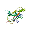
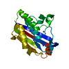
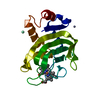
| Entry | Database: PDB / ID: 2f4w | ||||||
|---|---|---|---|---|---|---|---|
| Title | Human ubiquitin-conjugating enzyme E2 J2 | ||||||
 Components Components | ubiquitin-conjugating enzyme E2, J2 | ||||||
 Keywords Keywords | LIGASE / Endoplasmic reticulum / Ubl conjugation pathway / Structural Genomics Consortium (SGC) | ||||||
| Function / homology |  Function and homology information Function and homology informationprotein K6-linked ubiquitination / : / E2 ubiquitin-conjugating enzyme / Insertion of tail-anchored proteins into the endoplasmic reticulum membrane / ubiquitin conjugating enzyme activity / response to unfolded protein / ubiquitin ligase complex / ERAD pathway / protein polyubiquitination / Antigen processing: Ubiquitination & Proteasome degradation ...protein K6-linked ubiquitination / : / E2 ubiquitin-conjugating enzyme / Insertion of tail-anchored proteins into the endoplasmic reticulum membrane / ubiquitin conjugating enzyme activity / response to unfolded protein / ubiquitin ligase complex / ERAD pathway / protein polyubiquitination / Antigen processing: Ubiquitination & Proteasome degradation / E3 ubiquitin ligases ubiquitinate target proteins / ubiquitin-dependent protein catabolic process / ubiquitin protein ligase binding / endoplasmic reticulum membrane / endoplasmic reticulum / ATP binding / nucleus / cytosol Similarity search - Function | ||||||
| Biological species |  Homo sapiens (human) Homo sapiens (human) | ||||||
| Method |  X-RAY DIFFRACTION / SYNCHROTRON / MOLECULAR REPLACEMENT / Resolution: 2 Å X-RAY DIFFRACTION / SYNCHROTRON / MOLECULAR REPLACEMENT / Resolution: 2 Å | ||||||
 Authors Authors | Walker, J.R. / Avvakumov, G.V. / Xue, S. / Finerty Jr., P.J. / Newman, E.M. / Mackenzie, F. / Weigelt, J. / Sundstrom, M. / Arrowsmith, C. / Edwards, A. ...Walker, J.R. / Avvakumov, G.V. / Xue, S. / Finerty Jr., P.J. / Newman, E.M. / Mackenzie, F. / Weigelt, J. / Sundstrom, M. / Arrowsmith, C. / Edwards, A. / Bochkarev, A. / Dhe-Paganon, S. / Structural Genomics Consortium (SGC) | ||||||
 Citation Citation | Journal: Mol Cell Proteomics / Year: 2012 Title: A human ubiquitin conjugating enzyme (E2)-HECT E3 ligase structure-function screen. Authors: Sheng, Y. / Hong, J.H. / Doherty, R. / Srikumar, T. / Shloush, J. / Avvakumov, G.V. / Walker, J.R. / Xue, S. / Neculai, D. / Wan, J.W. / Kim, S.K. / Arrowsmith, C.H. / Raught, B. / Dhe-Paganon, S. #1: Journal: J.Biol.Chem. / Year: 2001 Title: Endoplasmic reticulum (ER)-associated degradation of T cell receptor subunits. Involvement of ER-associated ubiquitin-conjugating enzymes (E2s). Authors: Tiwari, S. / Weissman, A.M. | ||||||
| History |
|
- Structure visualization
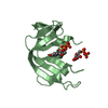
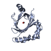
| Structure viewer | Molecule: MolmilJmol/JSmol |
|---|
- Downloads & links
-Download
| PDBx/mmCIF format | 2f4w.cif.gz | 79 KB | Display | PDBx/mmCIF format |
|---|---|---|---|---|
| PDB format | pdb2f4w.ent.gz | 58.5 KB | Display | PDB format |
| PDBx/mmJSON format | 2f4w.json.gz | Tree view | PDBx/mmJSON format | |
| Others |  Other downloads Other downloads |
-Validation report
| Arichive directory | https://data.pdbj.org/pub/pdb/validation_reports/f4/2f4wftp://data.pdbj.org/pub/pdb/validation_reports/f4/2f4w | HTTPS FTP |
|---|
-Related structure data
| Related structure data |  1y6lC 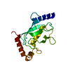 1yh2C  1yrvC 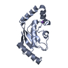 1zdnC  1zuoC 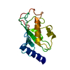 2a4dC  2a7lC  2awfC  2ob4C  2qgxC  2z5dC  3bzhC 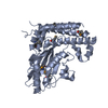 3cegC 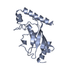 1ayzS S: Starting model for refinement C: citing same article ( |
|---|---|
| Similar structure data |


-Links
 PDBj
PDBj


- Assembly
| Deposited unit | 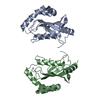
| ||||||||
|---|---|---|---|---|---|---|---|---|---|
| 1 | 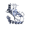
| ||||||||
| 2 | 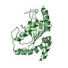
| ||||||||
| Unit cell |
| ||||||||
| Components on special symmetry positions |
|
-Components
| #1: Protein | Mass: 21332.525 Da / Num. of mol.: 2 Source method: isolated from a genetically manipulated source Source: (gene. exp.) Homo sapiens (human) / Gene: UBE2J2 / Plasmid: pET-28LIC / Species (production host): Escherichia coli / Production host:  References: GenBank: 56206319, UniProt: Q8N2K1*PLUS, ubiquitin-protein ligase #2: Water | ChemComp-HOH / |  Mass: 18.015 Da / Num. of mol.: 245 / Source method: isolated from a natural source / Formula: H2O Mass: 18.015 Da / Num. of mol.: 245 / Source method: isolated from a natural source / Formula: H2O |
|---|
-Experimental details
-Experiment
| Experiment | Method: X-RAY DIFFRACTION / Number of used crystals: 1 |
|---|
- Sample preparation
| Crystal | Density Matthews: 1.91 Å3/Da / Density % sol: 35.75 % |
|---|---|
| Crystal grow | Temperature: 298 K / Details: VAPOR DIFFUSION, HANGING DROP, temperature 298.0K |
-Data collection
| Diffraction | Mean temperature: 100 K |
|---|---|
| Diffraction source | Source: SYNCHROTRON / Site: APS  / Beamline: 19-BM / Wavelength: 1.00769 / Beamline: 19-BM / Wavelength: 1.00769 |
| Detector | Type: SBC-3 / Detector: CCD / Date: Nov 4, 2005 |
| Radiation | Protocol: SINGLE WAVELENGTH / Monochromatic (M) / Laue (L): M / Scattering type: x-ray |
| Radiation wavelength | Wavelength: 1.00769 Å / Relative weight: 1 |
| Reflection | Resolution: 2→30.84 Å / Num. obs: 23045 / % possible obs: 95.8 % / Observed criterion σ(I): -3 / Redundancy: 3.3 % / Rmerge(I) obs: 0.067 / Net I/σ(I): 23.67 |
| Reflection shell | Resolution: 2→2.07 Å / Redundancy: 2.9 % / Rmerge(I) obs: 0.238 / Mean I/σ(I) obs: 4.74 / % possible all: 85.6 |
- Processing
| Software |
| ||||||||||||||||||||||||||||||||||||||||||||||||||||||||||||||||||||||||||||||||||||||||||||||||||||||||||||||||||||||||||||||||||||||||||||||||||||||||||||||||||||||||||
|---|---|---|---|---|---|---|---|---|---|---|---|---|---|---|---|---|---|---|---|---|---|---|---|---|---|---|---|---|---|---|---|---|---|---|---|---|---|---|---|---|---|---|---|---|---|---|---|---|---|---|---|---|---|---|---|---|---|---|---|---|---|---|---|---|---|---|---|---|---|---|---|---|---|---|---|---|---|---|---|---|---|---|---|---|---|---|---|---|---|---|---|---|---|---|---|---|---|---|---|---|---|---|---|---|---|---|---|---|---|---|---|---|---|---|---|---|---|---|---|---|---|---|---|---|---|---|---|---|---|---|---|---|---|---|---|---|---|---|---|---|---|---|---|---|---|---|---|---|---|---|---|---|---|---|---|---|---|---|---|---|---|---|---|---|---|---|---|---|---|---|---|
| Refinement | Method to determine structure: MOLECULAR REPLACEMENT Starting model: 1AYZ Resolution: 2→30.84 Å / Cor.coef. Fo:Fc: 0.957 / Cor.coef. Fo:Fc free: 0.921 / SU B: 5.731 / SU ML: 0.158 / Cross valid method: THROUGHOUT / ESU R: 0.193 / ESU R Free: 0.183 / Stereochemistry target values: MAXIMUM LIKELIHOOD / Details: HYDROGENS HAVE BEEN ADDED IN THE RIDING POSITIONS
| ||||||||||||||||||||||||||||||||||||||||||||||||||||||||||||||||||||||||||||||||||||||||||||||||||||||||||||||||||||||||||||||||||||||||||||||||||||||||||||||||||||||||||
| Solvent computation | Ion probe radii: 0.8 Å / Shrinkage radii: 0.8 Å / VDW probe radii: 1.4 Å / Solvent model: BABINET MODEL WITH MASK | ||||||||||||||||||||||||||||||||||||||||||||||||||||||||||||||||||||||||||||||||||||||||||||||||||||||||||||||||||||||||||||||||||||||||||||||||||||||||||||||||||||||||||
| Displacement parameters | Biso mean: 36.137 Å2
| ||||||||||||||||||||||||||||||||||||||||||||||||||||||||||||||||||||||||||||||||||||||||||||||||||||||||||||||||||||||||||||||||||||||||||||||||||||||||||||||||||||||||||
| Refinement step | Cycle: LAST / Resolution: 2→30.84 Å
| ||||||||||||||||||||||||||||||||||||||||||||||||||||||||||||||||||||||||||||||||||||||||||||||||||||||||||||||||||||||||||||||||||||||||||||||||||||||||||||||||||||||||||
| Refine LS restraints |
| ||||||||||||||||||||||||||||||||||||||||||||||||||||||||||||||||||||||||||||||||||||||||||||||||||||||||||||||||||||||||||||||||||||||||||||||||||||||||||||||||||||||||||
| LS refinement shell | Resolution: 2→2.052 Å / Total num. of bins used: 20
|
