 Movie
Movie Controller

[English] 日本語
 Yorodumi
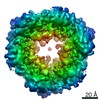
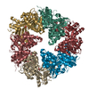
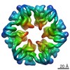
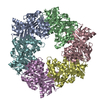
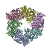
- PDB-2eww: Crystal Structure of the Pilus Retraction Motor PilT and Bound ATP -
+ Open data

- Basic information
| Entry | Database: PDB / ID: 2eww | ||||||
|---|---|---|---|---|---|---|---|
| Title | Crystal Structure of the Pilus Retraction Motor PilT and Bound ATP | ||||||
 Components Components | twitching motility protein PilT | ||||||
 Keywords Keywords | PROTEIN TRANSPORT / Pilus Retraction Motor / ATPase / Hexameric PilT | ||||||
| Function / homology |  Function and homology information Function and homology informationATP hydrolysis activity / ATP binding / identical protein binding / cytoplasm Similarity search - Function | ||||||
| Biological species |   Aquifex aeolicus (bacteria) Aquifex aeolicus (bacteria) | ||||||
| Method |  X-RAY DIFFRACTION / SYNCHROTRON / MAD / Resolution: 3.2 Å X-RAY DIFFRACTION / SYNCHROTRON / MAD / Resolution: 3.2 Å | ||||||
 Authors Authors | Satyshur, K.A. / Forest, K.T. | ||||||
 Citation Citation | Journal: Structure / Year: 2007 Title: Crystal structures of the pilus retraction motor PilT suggest large domain movements and subunit cooperation drive motility. Authors: Satyshur, K.A. / Worzalla, G.A. / Meyer, L.S. / Heiniger, E.K. / Aukema, K.G. / Misic, A.M. / Forest, K.T. #1: Journal: Acta Crystallogr.,Sect.D / Year: 2004 Title: The pilus-retraction protein PilT: ultrastructure of the biological assembly Authors: Forest, K.T. / Satyshur, K.A. / Worzalla, G.A. / Hansen, J.K. / Herdendorf, T.J. | ||||||
| History |
|
- Structure visualization
| Structure viewer | Molecule: MolmilJmol/JSmol |
|---|
- Downloads & links
-Download
| PDBx/mmCIF format | 2eww.cif.gz | 81.5 KB | Display | PDBx/mmCIF format |
|---|---|---|---|---|
| PDB format | pdb2eww.ent.gz | 61.1 KB | Display | PDB format |
| PDBx/mmJSON format | 2eww.json.gz | Tree view | PDBx/mmJSON format | |
| Others |  Other downloads Other downloads |
-Validation report
| Arichive directory | https://data.pdbj.org/pub/pdb/validation_reports/ew/2ewwftp://data.pdbj.org/pub/pdb/validation_reports/ew/2eww | HTTPS FTP |
|---|
-Related structure data


-Links
 PDBj
PDBj
- Assembly
| Deposited unit | 
| ||||||||
|---|---|---|---|---|---|---|---|---|---|
| 1 | x 6
| ||||||||
| Unit cell |
| ||||||||
| Details | The biological assembly is a hexamer generated from the monomer in the asymmetric unit by all six operations in P6. |
-Components
| #1: Protein | Mass: 42559.398 Da / Num. of mol.: 1 Source method: isolated from a genetically manipulated source Source: (gene. exp.) Aquifex aeolicus (bacteria) / Plasmid: pET23a / Production host: |
|---|---|
| #2: Chemical | ChemComp-ATP /   Mass: 507.181 Da / Num. of mol.: 1 / Source method: obtained synthetically / Formula: C10H16N5O13P3 / Comment: ATP, energy-carrying molecule*YM Mass: 507.181 Da / Num. of mol.: 1 / Source method: obtained synthetically / Formula: C10H16N5O13P3 / Comment: ATP, energy-carrying molecule*YM |
| #3: Water | ChemComp-HOH /  Mass: 18.015 Da / Num. of mol.: 2 / Source method: isolated from a natural source / Formula: H2O Mass: 18.015 Da / Num. of mol.: 2 / Source method: isolated from a natural source / Formula: H2O |
| Has protein modification | Y |
-Experimental details
-Experiment
| Experiment | Method: X-RAY DIFFRACTION / Number of used crystals: 1 |
|---|
- Sample preparation
| Crystal | Density Matthews: 2.69 Å3/Da / Density % sol: 54.31 % |
|---|---|
| Crystal grow | Temperature: 293 K / Method: vapor diffusion, hanging drop / pH: 7.9 Details: 5-8 mg/ml protein 30-45% MPD 0.2-0.4 M Ammonium Sulfate Tris, 15mM KCl 75mM 5% glycerol 5-10mM magnesium Chloride 1-5 mM ATP , pH 7.9, VAPOR DIFFUSION, HANGING DROP, temperature 293K |
-Data collection
| Diffraction |
| |||||||||||||||
|---|---|---|---|---|---|---|---|---|---|---|---|---|---|---|---|---|
| Diffraction source |
| |||||||||||||||
| Detector | Type: MARRESEARCH / Detector: CCD / Date: Nov 1, 2003 | |||||||||||||||
| Radiation | Monochromator: Diamond / Protocol: MAD / Monochromatic (M) / Laue (L): M / Scattering type: x-ray | |||||||||||||||
| Radiation wavelength |
| |||||||||||||||
| Reflection | Resolution: 3.2→20 Å / Num. obs: 7481 / % possible obs: 99.5 % / Observed criterion σ(F): 0 / Observed criterion σ(I): 0 / Rmerge(I) obs: 0.079 / Net I/σ(I): 7.9 | |||||||||||||||
| Reflection shell | Resolution: 3.2→3.37 Å / Rmerge(I) obs: 0.256 / Mean I/σ(I) obs: 2.9 / Num. unique all: 1093 / % possible all: 98 |

- Processing
| Software |
| ||||||||||||||||||||||||||||||||||||||||||||||||||||||||||||||||||||||||||||||||||||||||||
|---|---|---|---|---|---|---|---|---|---|---|---|---|---|---|---|---|---|---|---|---|---|---|---|---|---|---|---|---|---|---|---|---|---|---|---|---|---|---|---|---|---|---|---|---|---|---|---|---|---|---|---|---|---|---|---|---|---|---|---|---|---|---|---|---|---|---|---|---|---|---|---|---|---|---|---|---|---|---|---|---|---|---|---|---|---|---|---|---|---|---|---|
| Refinement | Method to determine structure: MAD / Resolution: 3.2→19.47 Å / Cor.coef. Fo:Fc: 0.914 / Cor.coef. Fo:Fc free: 0.838 / SU B: 29.41 / SU ML: 0.499 / Isotropic thermal model: isotropic / Cross valid method: THROUGHOUT / σ(F): 0 / ESU R Free: 0.642 / Stereochemistry target values: MAXIMUM LIKELIHOOD
| ||||||||||||||||||||||||||||||||||||||||||||||||||||||||||||||||||||||||||||||||||||||||||
| Solvent computation | Ion probe radii: 0.8 Å / Shrinkage radii: 0.8 Å / VDW probe radii: 1.2 Å / Solvent model: BABINET MODEL WITH MASK | ||||||||||||||||||||||||||||||||||||||||||||||||||||||||||||||||||||||||||||||||||||||||||
| Displacement parameters | Biso mean: 70.716 Å2
| ||||||||||||||||||||||||||||||||||||||||||||||||||||||||||||||||||||||||||||||||||||||||||
| Refinement step | Cycle: LAST / Resolution: 3.2→19.47 Å
| ||||||||||||||||||||||||||||||||||||||||||||||||||||||||||||||||||||||||||||||||||||||||||
| Refine LS restraints |
| ||||||||||||||||||||||||||||||||||||||||||||||||||||||||||||||||||||||||||||||||||||||||||
| LS refinement shell | Resolution: 3.203→3.284 Å / Rfactor Rfree error: 0.01 / Total num. of bins used: 20
|
