 Movie
Movie Controller

[English] 日本語
 Yorodumi
- PDB-2eyu: The Crystal Structure of the C-terminal Domain of Aquifex aeolicu... -
+ Open data

- Basic information
| Entry | Database: PDB / ID: 2eyu | ||||||
|---|---|---|---|---|---|---|---|
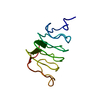
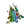
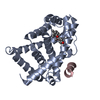
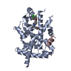
| Title | The Crystal Structure of the C-terminal Domain of Aquifex aeolicus PilT | ||||||
 Components Components | twitching motility protein PilT | ||||||
 Keywords Keywords | PROTEIN TRANSPORT / Pilus Retraction Motor / C-terminal Domain PilT | ||||||
| Function / homology |  Function and homology information Function and homology informationATP hydrolysis activity / ATP binding / identical protein binding / cytoplasm Similarity search - Function | ||||||
| Biological species |   Aquifex aeolicus (bacteria) Aquifex aeolicus (bacteria) | ||||||
| Method |  X-RAY DIFFRACTION / SYNCHROTRON / AB INITIO / Resolution: 1.87 Å X-RAY DIFFRACTION / SYNCHROTRON / AB INITIO / Resolution: 1.87 Å | ||||||
 Authors Authors | Satyshur, K.A. / Worzalla, G.A. / Meyer, L.S. / Heiniger, E.K. / Aukema, K.G. / Forest, K.T. | ||||||
 Citation Citation | Journal: Structure / Year: 2007 Title: Crystal structures of the pilus retraction motor PilT suggest large domain movements and subunit cooperation drive motility. Authors: Satyshur, K.A. / Worzalla, G.A. / Meyer, L.S. / Heiniger, E.K. / Aukema, K.G. / Misic, A.M. / Forest, K.T. #1: Journal: Acta Crystallogr.,Sect.D / Year: 2004 Title: The pilus-retraction protein PilT: ultrastructure of the biological assembly Authors: Forest, K.T. / Satyshur, K.A. / Worzalla, G.A. / Hansen, J.K. / Herdendorf, T.J. | ||||||
| History |
|
- Structure visualization
| Structure viewer | Molecule: MolmilJmol/JSmol |
|---|
- Downloads & links
-Download
| PDBx/mmCIF format | 2eyu.cif.gz | 109.8 KB | Display | PDBx/mmCIF format |
|---|---|---|---|---|
| PDB format | pdb2eyu.ent.gz | 84.8 KB | Display | PDB format |
| PDBx/mmJSON format | 2eyu.json.gz | Tree view | PDBx/mmJSON format | |
| Others |  Other downloads Other downloads |
-Validation report
| Arichive directory | https://data.pdbj.org/pub/pdb/validation_reports/ey/2eyuftp://data.pdbj.org/pub/pdb/validation_reports/ey/2eyu | HTTPS FTP |
|---|
-Related structure data


-Links
 PDBj
PDBj
- Assembly
| Deposited unit | 
| ||||||||
|---|---|---|---|---|---|---|---|---|---|
| 1 | 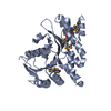
| ||||||||
| 2 | 
| ||||||||
| Unit cell |
|
-Components
| #1: Protein | Mass: 29873.084 Da / Num. of mol.: 2 / Fragment: PilT C-terminal Domain / Mutation: Glu294Gly Source method: isolated from a genetically manipulated source Source: (gene. exp.) Aquifex aeolicus (bacteria) / Plasmid: pET-23a / Production host: #2: Chemical | ChemComp-SO4 / |   Mass: 96.063 Da / Num. of mol.: 1 / Source method: obtained synthetically / Formula: SO4 Mass: 96.063 Da / Num. of mol.: 1 / Source method: obtained synthetically / Formula: SO4#3: Water | ChemComp-HOH / |  Mass: 18.015 Da / Num. of mol.: 17 / Source method: isolated from a natural source / Formula: H2O Mass: 18.015 Da / Num. of mol.: 17 / Source method: isolated from a natural source / Formula: H2OHas protein modification | Y | |
|---|
-Experimental details
-Experiment
| Experiment | Method: X-RAY DIFFRACTION / Number of used crystals: 1 |
|---|
- Sample preparation
| Crystal | Density Matthews: 2.17 Å3/Da / Density % sol: 47.59 % |
|---|---|
| Crystal grow | Temperature: 293 K / Method: vapor diffusion, hanging drop / pH: 6.5 Details: 10 mg/ml protein 0.1 M Mes 10% Dioxane 1.6 M ammonium sulfate , pH 6.5, VAPOR DIFFUSION, HANGING DROP, temperature 293K |
-Data collection
| Diffraction | Mean temperature: 100 K |
|---|---|
| Diffraction source | Source: SYNCHROTRON / Site: APS  / Beamline: 32-ID / Wavelength: 0.97934 Å / Beamline: 32-ID / Wavelength: 0.97934 Å |
| Detector | Type: MARRESEARCH / Detector: CCD / Date: Dec 11, 2004 |
| Radiation | Protocol: SINGLE WAVELENGTH / Monochromatic (M) / Laue (L): M / Scattering type: x-ray |
| Radiation wavelength | Wavelength: 0.97934 Å / Relative weight: 1 |
| Reflection | Resolution: 1.86→40 Å / Num. all: 45173 / Num. obs: 44351 / % possible obs: 96.8 % / Observed criterion σ(F): 4 / Observed criterion σ(I): 2 / Redundancy: 12 % / Rmerge(I) obs: 0.08 / Net I/σ(I): 49.9 |
| Reflection shell | Resolution: 1.86→1.94 Å / Redundancy: 6.9 % / Rmerge(I) obs: 0.197 / Mean I/σ(I) obs: 7.8 / % possible all: 83.9 |
- Processing
| Software |
| |||||||||||||||||||||||||||||||||
|---|---|---|---|---|---|---|---|---|---|---|---|---|---|---|---|---|---|---|---|---|---|---|---|---|---|---|---|---|---|---|---|---|---|---|
| Refinement | Method to determine structure: AB INITIO / Resolution: 1.87→20 Å / Num. parameters: 15692 / Num. restraintsaints: 16007 / Cross valid method: FREE R / σ(F): 4 / Stereochemistry target values: ENGH AND HUBER Details: Crystal is twinned, with BASF refined to 0.535 in ShelX h,-k,-l
| |||||||||||||||||||||||||||||||||
| Displacement parameters | Biso mean: 35.3 Å2 | |||||||||||||||||||||||||||||||||
| Refine analyze | Num. disordered residues: 0 / Occupancy sum hydrogen: 0 / Occupancy sum non hydrogen: 3922 | |||||||||||||||||||||||||||||||||
| Refinement step | Cycle: LAST / Resolution: 1.87→20 Å
| |||||||||||||||||||||||||||||||||
| Refine LS restraints |
| |||||||||||||||||||||||||||||||||
| LS refinement shell | Resolution: 1.87→1.871 Å /
|
