[English] 日本語
Yorodumi
- PDB-2gsz: Structure of A. aeolicus PilT with 6 monomers per asymmetric unit -

+
Open data


ID or keywords:

Loading...

-
Basic information

Entry
Database: PDB / ID: 2gsz
TitleStructure of A. aeolicus PilT with 6 monomers per asymmetric unit
Componentstwitching motility protein PilT
KeywordsPROTEIN TRANSPORT / P-loop / domain motion / ATPase / PAS / RecA
Function / homology
Function and homology information


ATP hydrolysis activity / ATP binding / identical protein binding
Similarity search - Function
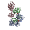
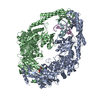
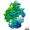
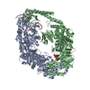
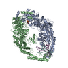
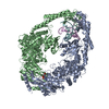
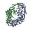
Pilus retraction protein PilT/PilU / : / Type II/IV secretion system protein / Type II/IV secretion system protein / ATPases associated with a variety of cellular activities / AAA+ ATPase domain / P-loop containing nucleoside triphosphate hydrolase
Similarity search - Domain/homology
ADENOSINE-5'-DIPHOSPHATE / : / Twitching motility protein PilT
Similarity search - Component
Biological speciesAquifex aeolicus (bacteria)
MethodX-RAY DIFFRACTION / SYNCHROTRON / MOLECULAR REPLACEMENT / Resolution: 4.2 Å
AuthorsForest, K.T. / Satyshur, K.A.
CitationJournal: Structure / Year: 2007
Title: Crystal structures of the pilus retraction motor PilT suggest large domain movements and subunit cooperation drive motility.
Authors: Satyshur, K.A. / Worzalla, G.A. / Meyer, L.S. / Heiniger, E.K. / Aukema, K.G. / Misic, A.M. / Forest, K.T.
History
DepositionApr 27, 2006Deposition site: RCSB / Processing site: RCSB
Revision 1.0Mar 20, 2007Provider: repository / Type: Initial release
Revision 1.1Oct 16, 2007Group: Version format compliance
Revision 1.2Jul 13, 2011Group: Version format compliance
Revision 1.3Oct 18, 2017Group: Refinement description / Category: software
Item: _software.classification / _software.contact_author ..._software.classification / _software.contact_author / _software.contact_author_email / _software.date / _software.language / _software.location / _software.name / _software.type / _software.version
Revision 1.4Aug 30, 2023Group: Data collection / Database references ...Data collection / Database references / Derived calculations / Refinement description
Category: chem_comp_atom / chem_comp_bond ...chem_comp_atom / chem_comp_bond / database_2 / pdbx_initial_refinement_model / struct_conn / struct_ncs_dom_lim / struct_ref_seq_dif / struct_site
Item: _database_2.pdbx_DOI / _database_2.pdbx_database_accession ..._database_2.pdbx_DOI / _database_2.pdbx_database_accession / _struct_conn.pdbx_leaving_atom_flag / _struct_ncs_dom_lim.beg_auth_comp_id / _struct_ncs_dom_lim.end_auth_comp_id / _struct_ref_seq_dif.details / _struct_site.pdbx_auth_asym_id / _struct_site.pdbx_auth_comp_id / _struct_site.pdbx_auth_seq_id
Revision 1.5Nov 15, 2023Group: Data collection / Category: chem_comp_atom / chem_comp_bond / Item: _chem_comp_atom.atom_id / _chem_comp_bond.atom_id_2

-
Structure visualization

Structure viewerMolecule:
MolmilJmol/JSmol

Downloads & links

-
Assembly

Deposited unit
A: twitching motility protein PilT
B: twitching motility protein PilT
C: twitching motility protein PilT
D: twitching motility protein PilT
E: twitching motility protein PilT
F: twitching motility protein PilT
hetero molecules


Theoretical massNumber of molelcules
Total (without water)248,79210
Polymers248,0776
Non-polymers7154
Water00
1


  • Idetical with deposited unit
  • defined by author
TypeNameSymmetry operationNumber
identity operation1_555x,y,z1
Unit cell
Length a, b, c (Å)206.111, 105.244, 123.023
Angle α, β, γ (deg.)90.00, 111.06, 90.00
Int Tables number5
Space group name H-MC121
Noncrystallographic symmetry (NCS)NCS domain:
IDEns-IDDetails
11A
21B
31C
41D
51E
61F
12A
22B
32C
42D
52E
62F

NCS domain segments:

Component-ID: 1 / Refine code: 1

Dom-IDEns-IDBeg auth comp-IDBeg label comp-IDEnd auth comp-IDEnd label comp-IDAuth asym-IDLabel asym-IDAuth seq-IDLabel seq-ID
11GLUGLUARGARGAA12 - 1101 - 99
21GLUGLUARGARGBB12 - 1101 - 99
31GLUGLUARGARGCC12 - 1101 - 99
41GLUGLUARGARGDD12 - 1101 - 99
51GLUGLUARGARGEE12 - 1101 - 99
61GLUGLUARGARGFF12 - 1101 - 99
12ILEILEMSEMSEAA116 - 361105 - 350
22ILEILEMSEMSEBB116 - 361105 - 350
32ILEILEMSEMSECC116 - 361105 - 350
42ILEILEMSEMSEDD116 - 361105 - 350
52ILEILEMSEMSEEE116 - 361105 - 350
62ILEILEMSEMSEFF116 - 361105 - 350

NCS ensembles :
ID
1
2
DetailsEach asymmetric unit contains one complete hexamer, the presumed biological assembly.

-
Components

#1: Protein
twitching motility protein PilT


Mass: 41346.145 Da / Num. of mol.: 6
Source method: isolated from a genetically manipulated source
Source: (gene. exp.) Aquifex aeolicus (bacteria) / Gene: pilT / Plasmid: pET23a+ / Species (production host): Escherichia coli / Production host: Escherichia coli BL21(DE3) (bacteria) / Strain (production host): BL21DE3 / References: GenBank: 2983313, UniProt: O66950*PLUS
#2: Chemical ChemComp-SO4 / SULFATE ION


Mass: 96.063 Da / Num. of mol.: 3 / Source method: obtained synthetically / Formula: SO4
#3: Chemical ChemComp-ADP / ADENOSINE-5'-DIPHOSPHATE


Mass: 427.201 Da / Num. of mol.: 1 / Source method: obtained synthetically / Formula: C10H15N5O10P2 / Comment: ADP, energy-carrying molecule*YM

-
Experimental details

-
Experiment

ExperimentMethod: X-RAY DIFFRACTION / Number of used crystals: 1

-
Sample preparation

CrystalDensity Matthews: 2.51 Å3/Da / Density % sol: 50.96 %
Crystal growTemperature: 293 K / Method: vapor diffusion, hanging drop / pH: 7.6
Details: Upon set up, protein droplet contains 7.5 mg/ml protein in 9.7% PEG1000, 13.0 mM TCEP, 13.0 mM LiCl, 0.32 M AmSO4, 2.6 mM ADP, 16 mM Tris pH 7.6, 98 mM KCl, 130 mM Imidazole, 6.5% glycerol. ...Details: Upon set up, protein droplet contains 7.5 mg/ml protein in 9.7% PEG1000, 13.0 mM TCEP, 13.0 mM LiCl, 0.32 M AmSO4, 2.6 mM ADP, 16 mM Tris pH 7.6, 98 mM KCl, 130 mM Imidazole, 6.5% glycerol. Well contains 0.2 M AmSO4, 10% PEG 1000, unbuffered., VAPOR DIFFUSION, HANGING DROP, temperature 293K

-
Data collection

DiffractionMean temperature: 100 K
Diffraction sourceSource: SYNCHROTRON / Site: APS / Beamline: 14-ID-B / Wavelength: 0.97896 Å
DetectorType: MAR CCD 165 mm / Detector: CCD / Date: Aug 2, 2004
RadiationProtocol: SINGLE WAVELENGTH / Monochromatic (M) / Laue (L): M / Scattering type: x-ray
Radiation wavelengthWavelength: 0.97896 Å / Relative weight: 1
ReflectionRedundancy: 3 % / Av σ(I) over netI: 9.1 / Number: 106618 / Rmerge(I) obs: 0.078 / Χ2: 1.05 / D res high: 4.1 Å / D res low: 50 Å / Num. obs: 35971 / % possible obs: 95.7
Diffraction reflection shell

ID: 1

Highest resolution (Å)Lowest resolution (Å)Num. obs% possible obs (%)Rmerge(I) obsChi squaredRedundancy
8.8250326086.90.0281.72.8
7.018.82359095.60.0411.4063
6.127.013654960.0720.9383
5.566.12361496.50.10.8883
5.165.56363796.40.1020.9163
4.865.16362596.90.1030.93
4.624.86363496.90.1170.9163
4.424.623661970.1641.0143
4.254.42359197.20.2221.1282.9
4.14.25370597.20.6220.8453
ReflectionResolution: 4.2→50 Å / Num. obs: 17281 / % possible obs: 95.2 % / Observed criterion σ(F): 0 / Observed criterion σ(I): 0 / Redundancy: 5.7 % / Rsym value: 0.089 / Net I/σ(I): 15.6
Reflection shellResolution: 4.2→4.35 Å / Redundancy: 5.8 % / Mean I/σ(I) obs: 5.3 / Rsym value: 0.276 / % possible all: 96.6

-
Processing

Software
NameVersionClassificationNB
DENZOdata reduction
SCALEPACKdata scaling
PHASERphasing
REFMACrefinement
PDB_EXTRACT1.701data extraction
ADSCdata collection
RefinementMethod to determine structure: MOLECULAR REPLACEMENT
Starting model: NTD of mol C and CTD of mol D from 2EWV.pdb
Resolution: 4.2→25 Å / Cor.coef. Fo:Fc: 0.822 / Cor.coef. Fo:Fc free: 0.768 / SU B: 129.953 / SU ML: 1.66 / Cross valid method: THROUGHOUT / ESU R Free: 1.77 / Stereochemistry target values: MAXIMUM LIKELIHOOD
RfactorNum. reflection% reflectionSelection details
Rfree0.40563 859 5 %RANDOM
Rwork0.34307 ---
obs0.34634 16243 94.79 %-
all-18247 --
Solvent computationIon probe radii: 0.8 Å / Shrinkage radii: 0.8 Å / VDW probe radii: 1.2 Å / Solvent model: BABINET MODEL WITH MASK
Displacement parametersBiso mean: 124.351 Å2
Baniso -1Baniso -2Baniso -3
1--6.13 Å20 Å2-7.32 Å2
2---0.19 Å20 Å2
3---1.06 Å2
Refinement stepCycle: LAST / Resolution: 4.2→25 Å
ProteinNucleic acidLigandSolventTotal
Num. atoms16302 0 42 0 16344
Refine LS restraints
Refine-IDTypeDev idealDev ideal targetNumber
X-RAY DIFFRACTIONr_bond_refined_d0.0190.02216613
X-RAY DIFFRACTIONr_bond_other_d
X-RAY DIFFRACTIONr_angle_refined_deg1.7321.98722431
X-RAY DIFFRACTIONr_angle_other_deg
X-RAY DIFFRACTIONr_dihedral_angle_1_deg7.74652046
X-RAY DIFFRACTIONr_dihedral_angle_2_deg38.95624.065738
X-RAY DIFFRACTIONr_dihedral_angle_3_deg25.761153126
X-RAY DIFFRACTIONr_dihedral_angle_4_deg21.93815126
X-RAY DIFFRACTIONr_chiral_restr0.0990.22596
X-RAY DIFFRACTIONr_gen_planes_refined0.0050.0212227
X-RAY DIFFRACTIONr_gen_planes_other
X-RAY DIFFRACTIONr_nbd_refined0.2780.27078
X-RAY DIFFRACTIONr_nbd_other
X-RAY DIFFRACTIONr_nbtor_refined0.3220.210833
X-RAY DIFFRACTIONr_nbtor_other
X-RAY DIFFRACTIONr_xyhbond_nbd_refined0.2030.2483
X-RAY DIFFRACTIONr_xyhbond_nbd_other
X-RAY DIFFRACTIONr_metal_ion_refined
X-RAY DIFFRACTIONr_metal_ion_other
X-RAY DIFFRACTIONr_symmetry_vdw_refined0.6080.292
X-RAY DIFFRACTIONr_symmetry_vdw_other
X-RAY DIFFRACTIONr_symmetry_hbond_refined0.6770.27
X-RAY DIFFRACTIONr_symmetry_hbond_other
X-RAY DIFFRACTIONr_symmetry_metal_ion_refined
X-RAY DIFFRACTIONr_symmetry_metal_ion_other
X-RAY DIFFRACTIONr_mcbond_it0.5261.510535
X-RAY DIFFRACTIONr_mcbond_other
X-RAY DIFFRACTIONr_mcangle_it0.881216608
X-RAY DIFFRACTIONr_scbond_it0.84536726
X-RAY DIFFRACTIONr_scangle_it1.3924.55823
X-RAY DIFFRACTIONr_rigid_bond_restr
X-RAY DIFFRACTIONr_sphericity_free
X-RAY DIFFRACTIONr_sphericity_bonded
Refine LS restraints NCS

Refine-ID: X-RAY DIFFRACTION

Ens-IDDom-IDAuth asym-IDNumberTypeRms dev position (Å)Weight position
11A783tight positional0.070.05
12B783tight positional0.070.05
13C783tight positional0.060.05
14D783tight positional0.070.05
15E783tight positional0.080.05
16F783tight positional0.060.05
21A1900tight positional0.070.05
22B1900tight positional0.070.05
23C1900tight positional0.060.05
24D1900tight positional0.070.05
25E1900tight positional0.070.05
26F1900tight positional0.070.05
11A783tight thermal0.110.5
12B783tight thermal0.080.5
13C783tight thermal0.080.5
14D783tight thermal0.090.5
15E783tight thermal0.080.5
16F783tight thermal0.070.5
21A1900tight thermal0.080.5
22B1900tight thermal0.070.5
23C1900tight thermal0.070.5
24D1900tight thermal0.080.5
25E1900tight thermal0.070.5
26F1900tight thermal0.080.5
LS refinement shellResolution: 4.2→4.306 Å / Total num. of bins used: 20
RfactorNum. reflection% reflection
Rfree0.408 53 -
Rwork0.418 1085 -
obs--87.4 %

+
About Yorodumi

-
News

-
Feb 9, 2022. New format data for meta-information of EMDB entries

New format data for meta-information of EMDB entries

  • Version 3 of the EMDB header file is now the official format.
  • The previous official version 1.9 will be removed from the archive.

Related info.:EMDB header

External links:wwPDB to switch to version 3 of the EMDB data model

-
Aug 12, 2020. Covid-19 info

Covid-19 info

URL: https://pdbjlc1.pdbj.org/emnavi/covid19.php

New page: Covid-19 featured information page in EM Navigator.

Related info.:Covid-19 info / Mar 5, 2020. Novel coronavirus structure data

+
Mar 5, 2020. Novel coronavirus structure data

Novel coronavirus structure data

Related info.:Yorodumi Speices / Aug 12, 2020. Covid-19 info

External links:COVID-19 featured content - PDBj / Molecule of the Month (242):Coronavirus Proteases

+
Jan 31, 2019. EMDB accession codes are about to change! (news from PDBe EMDB page)

EMDB accession codes are about to change! (news from PDBe EMDB page)

  • The allocation of 4 digits for EMDB accession codes will soon come to an end. Whilst these codes will remain in use, new EMDB accession codes will include an additional digit and will expand incrementally as the available range of codes is exhausted. The current 4-digit format prefixed with “EMD-” (i.e. EMD-XXXX) will advance to a 5-digit format (i.e. EMD-XXXXX), and so on. It is currently estimated that the 4-digit codes will be depleted around Spring 2019, at which point the 5-digit format will come into force.
  • The EM Navigator/Yorodumi systems omit the EMD- prefix.

Related info.:Q: What is EMD? / ID/Accession-code notation in Yorodumi/EM Navigator

External links:EMDB Accession Codes are Changing Soon! / Contact to PDBj

+
Jul 12, 2017. Major update of PDB

Major update of PDB

  • wwPDB released updated PDB data conforming to the new PDBx/mmCIF dictionary.
  • This is a major update changing the version number from 4 to 5, and with Remediation, in which all the entries are updated.
  • In this update, many items about electron microscopy experimental information are reorganized (e.g. em_software).
  • Now, EM Navigator and Yorodumi are based on the updated data.

External links:wwPDB Remediation / Enriched Model Files Conforming to OneDep Data Standards Now Available in the PDB FTP Archive

-
Yorodumi

Thousand views of thousand structures

  • Yorodumi is a browser for structure data from EMDB, PDB, SASBDB, etc.
  • This page is also the successor to EM Navigator detail page, and also detail information page/front-end page for Omokage search.
  • The word "yorodu" (or yorozu) is an old Japanese word meaning "ten thousand". "mi" (miru) is to see.

Related info.:EMDB / PDB / SASBDB / Comparison of 3 databanks / Yorodumi Search / Aug 31, 2016. New EM Navigator & Yorodumi / Yorodumi Papers / Jmol/JSmol / Function and homology information / Changes in new EM Navigator and Yorodumi

Read more