 Movie
Movie Controller

[English] 日本語
 Yorodumi
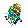
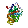
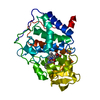
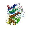
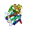
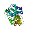
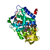
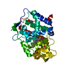
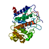
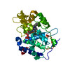
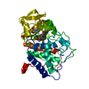
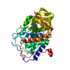
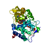
- PDB-2cyp: Crystal structure of yeast cytochrome C peroxidase refined at 1.7... -
+ Open data

- Basic information
| Entry | Database: PDB / ID: 2cyp | |||||||||
|---|---|---|---|---|---|---|---|---|---|---|
| Title | Crystal structure of yeast cytochrome C peroxidase refined at 1.7-angstroms resolution | |||||||||
 Components Components | CYTOCHROME C PEROXIDASE | |||||||||
 Keywords Keywords | OXIDOREDUCTASE (H2O2(A)) | |||||||||
| Function / homology |  Function and homology informationcytochrome-c peroxidase / cytochrome-c peroxidase activity / response to reactive oxygen species / hydrogen peroxide catabolic process / peroxidase activity / mitochondrial intermembrane space / cellular response to oxidative stress / mitochondrial matrix / heme binding / mitochondrion / metal ion binding Function and homology informationcytochrome-c peroxidase / cytochrome-c peroxidase activity / response to reactive oxygen species / hydrogen peroxide catabolic process / peroxidase activity / mitochondrial intermembrane space / cellular response to oxidative stress / mitochondrial matrix / heme binding / mitochondrion / metal ion bindingSimilarity search - Function | |||||||||
| Biological species |  Saccharomyces cerevisiae (brewer's yeast) Saccharomyces cerevisiae (brewer's yeast) | |||||||||
| Method | X-RAY DIFFRACTION / Resolution: 1.7 Å | |||||||||
 Authors Authors | Finzel, B.C. / Poulos, T.L. / Kraut, J. | |||||||||
 Citation Citation | Journal: J.Biol.Chem. / Year: 1984 Title: Crystal structure of yeast cytochrome c peroxidase refined at 1.7-A resolution Authors: Finzel, B.C. / Poulos, T.L. / Kraut, J. #1: Journal: Pept.Protein Rev. / Year: 1984Title: Heme Enzyme Structure and Function Authors: Poulos, T.L. / Finzel, B.C. #2: Journal: J.Biol.Chem. / Year: 1980Title: A Hypothetical Model of the Cytochrome C Peroxidase-Cytochrome C Electron Transfer Complex Authors: Poulos, T.L. / Kraut, J. #3: Journal: J.Biol.Chem. / Year: 1980Title: The Stereochemistry of Peroxidase Catalysis Authors: Poulos, T.L. / Kraut, J. #4: Journal: J.Biol.Chem. / Year: 1980Title: The Crystal Structure of Cytochrome C Peroxidase Authors: Poulos, T.L. / Freer, S.T. / Alden, R.A. / Edwards, S.L. / Skogland, U. / Takio, K. / Eriksson, B. / Xuong, N.-H. / Yonetani, T. / Kraut, J. #5: Journal: J.Biol.Chem. / Year: 1978Title: Crystallographic Determination of the Heme Orientation and Location of the Cyanide Binding Site in Yeast Cytochrome C Peroxidase Authors: Poulos, T.L. / Freer, S.T. / Alden, R.A. / Xuong, N.H. / Edwards, S.L. / Hamlin, R.C. / Kraut, J. | |||||||||
| History |
|
- Structure visualization
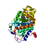
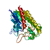
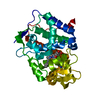
| Structure viewer | Molecule: MolmilJmol/JSmol |
|---|
- Downloads & links
-Download
| PDBx/mmCIF format | 2cyp.cif.gz | 79.4 KB | Display | PDBx/mmCIF format |
|---|---|---|---|---|
| PDB format | pdb2cyp.ent.gz | 58.4 KB | Display | PDB format |
| PDBx/mmJSON format | 2cyp.json.gz | Tree view | PDBx/mmJSON format | |
| Others |  Other downloads Other downloads |
-Validation report
| Arichive directory | https://data.pdbj.org/pub/pdb/validation_reports/cy/2cypftp://data.pdbj.org/pub/pdb/validation_reports/cy/2cyp | HTTPS FTP |
|---|
-Related structure data
| Similar structure data |
|---|


-Links
 PDBj
PDBj


- Assembly
| Deposited unit | 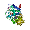
| ||||||||
|---|---|---|---|---|---|---|---|---|---|
| 1 |
| ||||||||
| Unit cell |
| ||||||||
| Atom site foot note | 1: THIS ATOM WAS DESIGNATED HOH 595 BY THE DEPOSITORS. / 2: SEE REMARK 4. |
-Components
| #1: Protein | Mass: 33571.238 Da / Num. of mol.: 1 Source method: isolated from a genetically manipulated source Source: (gene. exp.) Saccharomyces cerevisiae (brewer's yeast)References: UniProt: P00431, cytochrome-c peroxidase |
|---|---|
| #2: Chemical | ChemComp-HEM / Heme B  Mass: 616.487 Da / Num. of mol.: 1 / Source method: obtained synthetically / Formula: C34H32FeN4O4 Mass: 616.487 Da / Num. of mol.: 1 / Source method: obtained synthetically / Formula: C34H32FeN4O4 |
| #3: Chemical | ChemComp-O / Oxygen  Mass: 15.999 Da / Num. of mol.: 1 / Source method: obtained synthetically / Formula: O Mass: 15.999 Da / Num. of mol.: 1 / Source method: obtained synthetically / Formula: O |
| #4: Water | ChemComp-HOH / Water Mass: 18.015 Da / Num. of mol.: 262 / Source method: isolated from a natural source / Formula: H2O Mass: 18.015 Da / Num. of mol.: 262 / Source method: isolated from a natural source / Formula: H2O |
-Experimental details
-Experiment
| Experiment | Method: X-RAY DIFFRACTION |
|---|
- Sample preparation
| Crystal | Density Matthews: 3.16 Å3/Da / Density % sol: 61.02 % | |||||||||||||||
|---|---|---|---|---|---|---|---|---|---|---|---|---|---|---|---|---|
| Crystal grow | *PLUS pH: 6 / Method: unknown | |||||||||||||||
| Components of the solutions | *PLUS
|
-Data collection
| Reflection | *PLUS Highest resolution: 1.7 Å / Num. obs: 44888 / Num. measured all: 181721 / Rmerge(I) obs: 0.07 |
|---|
- Processing
| Software | Name: PROLSQ / Classification: refinement | ||||||||||||||||||||||||||||||||||||||||||||||||||||||||||||
|---|---|---|---|---|---|---|---|---|---|---|---|---|---|---|---|---|---|---|---|---|---|---|---|---|---|---|---|---|---|---|---|---|---|---|---|---|---|---|---|---|---|---|---|---|---|---|---|---|---|---|---|---|---|---|---|---|---|---|---|---|---|
| Refinement | Rfactor Rwork: 0.202 / Highest resolution: 1.7 Å | ||||||||||||||||||||||||||||||||||||||||||||||||||||||||||||
| Refinement step | Cycle: LAST / Highest resolution: 1.7 Å
| ||||||||||||||||||||||||||||||||||||||||||||||||||||||||||||
| Refine LS restraints |
| ||||||||||||||||||||||||||||||||||||||||||||||||||||||||||||
| Refinement | *PLUS Num. reflection obs: 44888 / Rfactor obs: 0.202 | ||||||||||||||||||||||||||||||||||||||||||||||||||||||||||||
| Solvent computation | *PLUS | ||||||||||||||||||||||||||||||||||||||||||||||||||||||||||||
| Displacement parameters | *PLUS | ||||||||||||||||||||||||||||||||||||||||||||||||||||||||||||
| Refine LS restraints | *PLUS Type: o_bond_d / Dev ideal target: 0.03 |
