 Movie
Movie Controller


+ Open data

- Basic information
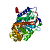
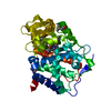
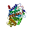
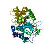
| Entry | Database: PDB / ID: 2as4 | ||||||
|---|---|---|---|---|---|---|---|
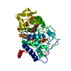
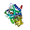
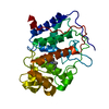
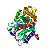
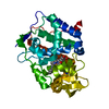
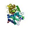
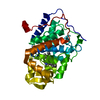
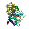
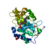
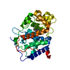
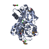
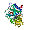
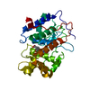
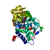
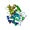
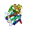
| Title | cytochrome c peroxidase in complex with 3-fluorocatechol | ||||||
 Components Components | Cytochrome c peroxidase, mitochondrial | ||||||
 Keywords Keywords | OXIDOREDUCTASE / peroxidase / model binding site | ||||||
| Function / homology |  Function and homology information Function and homology informationcytochrome-c peroxidase / cytochrome-c peroxidase activity / hydrogen peroxide catabolic process / response to reactive oxygen species / peroxidase activity / mitochondrial intermembrane space / cellular response to oxidative stress / mitochondrial matrix / heme binding / mitochondrion / metal ion binding Similarity search - Function | ||||||
| Biological species |  | ||||||
| Method |  X-RAY DIFFRACTION / SYNCHROTRON / FOURIER SYNTHESIS / Resolution: 1.3 Å X-RAY DIFFRACTION / SYNCHROTRON / FOURIER SYNTHESIS / Resolution: 1.3 Å | ||||||
 Authors Authors | Brenk, R. / Vetter, S.W. / Boyce, S.E. / Goodin, D.B. / Shoichet, B.K. | ||||||
 Citation Citation | Journal: J.Mol.Biol. / Year: 2006 Title: Probing molecular docking in a charged model binding site. Authors: Brenk, R. / Vetter, S.W. / Boyce, S.E. / Goodin, D.B. / Shoichet, B.K. | ||||||
| History |
|
- Structure visualization
| Structure viewer | Molecule: MolmilJmol/JSmol |
|---|
- Downloads & links
-Download
| PDBx/mmCIF format | 2as4.cif.gz | 149.9 KB | Display | PDBx/mmCIF format |
|---|---|---|---|---|
| PDB format | pdb2as4.ent.gz | 114.8 KB | Display | PDB format |
| PDBx/mmJSON format | 2as4.json.gz | Tree view | PDBx/mmJSON format | |
| Others |  Other downloads Other downloads |
-Validation report
| Arichive directory | https://data.pdbj.org/pub/pdb/validation_reports/as/2as4ftp://data.pdbj.org/pub/pdb/validation_reports/as/2as4 | HTTPS FTP |
|---|
-Related structure data
| Related structure data |  2anzC  2aqdC  2as1C  2as2C 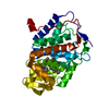 2as3C  2as6C 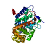 2eunC  2euoC  2eupC 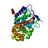 2euqC  2eurC  2eusC  2eutC  2euuC 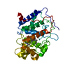 1ac4S C: citing same article ( S: Starting model for refinement |
|---|---|
| Similar structure data |

-Links
 PDBj
PDBj
- Assembly
| Deposited unit | 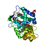
| ||||||||
|---|---|---|---|---|---|---|---|---|---|
| 1 |
| ||||||||
| Unit cell |
|
-Components
| #1: Protein | Mass: 33458.258 Da / Num. of mol.: 1 / Fragment: cytochrome c peroxidase / Mutation: W191G Source method: isolated from a genetically manipulated source Source: (gene. exp.) Gene: CCP1, CCP, CPO / Plasmid: PT7CCP / Species (production host): Escherichia coli / Production host:  | ||
|---|---|---|---|
| #2: Chemical | ChemComp-K /   Mass: 39.098 Da / Num. of mol.: 1 / Source method: obtained synthetically / Formula: K Mass: 39.098 Da / Num. of mol.: 1 / Source method: obtained synthetically / Formula: K | ||
| #3: Chemical | ChemComp-HEM /   Mass: 616.487 Da / Num. of mol.: 1 / Source method: obtained synthetically / Formula: C34H32FeN4O4 Mass: 616.487 Da / Num. of mol.: 1 / Source method: obtained synthetically / Formula: C34H32FeN4O4 | ||
| #4: Chemical |   Mass: 128.101 Da / Num. of mol.: 2 / Source method: obtained synthetically / Formula: C6H5FO2 Mass: 128.101 Da / Num. of mol.: 2 / Source method: obtained synthetically / Formula: C6H5FO2#5: Water | ChemComp-HOH / |  Mass: 18.015 Da / Num. of mol.: 375 / Source method: isolated from a natural source / Formula: H2O Mass: 18.015 Da / Num. of mol.: 375 / Source method: isolated from a natural source / Formula: H2O |
-Experimental details
-Experiment
| Experiment | Method: X-RAY DIFFRACTION / Number of used crystals: 1 |
|---|
- Sample preparation
| Crystal | Density Matthews: 3.9 Å3/Da / Density % sol: 59.99 % |
|---|---|
| Crystal grow | Temperature: 291 K / Method: vapor diffusion, hanging drop / pH: 4.5 Details: MPD, acetate, pH 4.5, VAPOR DIFFUSION, HANGING DROP, temperature 291K |
-Data collection
| Diffraction | Mean temperature: 100 K |
|---|---|
| Diffraction source | Source: SYNCHROTRON / Site: ALS  / Beamline: 8.3.1 / Wavelength: 1.11588 Å / Beamline: 8.3.1 / Wavelength: 1.11588 Å |
| Detector | Type: ADSC QUANTUM 4 / Detector: CCD / Date: Apr 10, 2005 |
| Radiation | Monochromator: double crystal / Protocol: SINGLE WAVELENGTH / Monochromatic (M) / Laue (L): M / Scattering type: x-ray |
| Radiation wavelength | Wavelength: 1.11588 Å / Relative weight: 1 |
| Reflection | Resolution: 1.3→10 Å / Num. all: 95083 / Num. obs: 95083 / % possible obs: 97 % / Rmerge(I) obs: 0.039 / Net I/σ(I): 31.3 |
| Reflection shell | Resolution: 1.3→1.35 Å / % possible all: 78.9 |
- Processing
| Software |
| |||||||||||||||||||||||||||||||||
|---|---|---|---|---|---|---|---|---|---|---|---|---|---|---|---|---|---|---|---|---|---|---|---|---|---|---|---|---|---|---|---|---|---|---|
| Refinement | Method to determine structure: FOURIER SYNTHESIS Starting model: 1AC4 Resolution: 1.3→10 Å / Num. parameters: 24731 / Num. restraintsaints: 30076 / Cross valid method: FREE R / σ(F): 0 / Stereochemistry target values: ENGH AND HUBER Details: ANISOTROPIC REFINEMENT REDUCED FREE R (NO CUTOFF) BY 7 % and the R-factor by 8 %
| |||||||||||||||||||||||||||||||||
| Refine analyze | Num. disordered residues: 10 / Occupancy sum hydrogen: 0 / Occupancy sum non hydrogen: 2726.91 | |||||||||||||||||||||||||||||||||
| Refinement step | Cycle: LAST / Resolution: 1.3→10 Å
| |||||||||||||||||||||||||||||||||
| Refine LS restraints |
|
