 Movie
Movie Controller

[English] 日本語
 Yorodumi
- PDB-2cfl: AGAO in complex with wc6b (Ru-wire inhibitor, 6-carbon linker, da... -
+ Open data

- Basic information
| Entry | Database: PDB / ID: 2cfl | ||||||
|---|---|---|---|---|---|---|---|
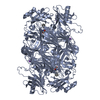
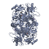
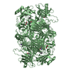
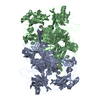
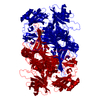
| Title | AGAO in complex with wc6b (Ru-wire inhibitor, 6-carbon linker, data set b) | ||||||
 Components Components | PHENYLETHYLAMINE OXIDASE | ||||||
 Keywords Keywords |  OXIDOREDUCTASE / AMINE OXIDASE / ARTHROBACTER GLOBIFORMIS / COPPER / COPPER CONTAINING / TPQ / CUAO / QUINONE / RUTHENIUM DIIMINE WIRES / COMPETITIVE INHIBITION / METAL-BINDING OXIDOREDUCTASE / AMINE OXIDASE / ARTHROBACTER GLOBIFORMIS / COPPER / COPPER CONTAINING / TPQ / CUAO / QUINONE / RUTHENIUM DIIMINE WIRES / COMPETITIVE INHIBITION / METAL-BINDING | ||||||
| Function / homology |  Function and homology information Function and homology information: / : / : / primary-amine oxidase / amine metabolic process / quinone binding / copper ion bindingSimilarity search - Function | ||||||
| Biological species |  ARTHROBACTER GLOBIFORMIS (bacteria) ARTHROBACTER GLOBIFORMIS (bacteria) | ||||||
| Method | X-RAY DIFFRACTION / MOLECULAR REPLACEMENT / Resolution: 1.8 Å | ||||||
 Authors Authors | Langley, D.B. / Duff, A.P. / Freeman, H.C. / Guss, J.M. / Juda, G.A. / Dooley, D.M. / Contakes, S.M. / Halpern-Manners, N.W. / Dunn, A.R. / Gray, H.B. | ||||||
 Citation Citation | Journal: J.Am.Chem.Soc. / Year: 2008 Title: Enantiomer-Specific Binding of Ruthenium(II) Molecular Wires by the Amine Oxidase of Arthrobacter Globiformis. Authors: Langley, D.B. / Brown, D.E. / Cheruzel, L.E. / Contakes, S.M. / Duff, A.P. / Hilmer, K.M. / Dooley, D.M. / Gray, H.B. / Guss, J.M. / Freeman, H.C. | ||||||
| History |
| ||||||
| Remark 700 | SHEET THE SHEET STRUCTURE OF THIS MOLECULE IS BIFURCATED. IN ORDER TO REPRESENT THIS FEATURE IN ... SHEET THE SHEET STRUCTURE OF THIS MOLECULE IS BIFURCATED. IN ORDER TO REPRESENT THIS FEATURE IN THE SHEET RECORDS BELOW, TWO SHEETS ARE DEFINED. |
- Structure visualization
| Structure viewer | Molecule: MolmilJmol/JSmol |
|---|
- Downloads & links
-Download
| PDBx/mmCIF format | 2cfl.cif.gz | 241.2 KB | Display | PDBx/mmCIF format |
|---|---|---|---|---|
| PDB format | pdb2cfl.ent.gz | 204.2 KB | Display | PDB format |
| PDBx/mmJSON format | 2cfl.json.gz | Tree view | PDBx/mmJSON format | |
| Others |  Other downloads Other downloads |
-Validation report
| Arichive directory | https://data.pdbj.org/pub/pdb/validation_reports/cf/2cflftp://data.pdbj.org/pub/pdb/validation_reports/cf/2cfl | HTTPS FTP |
|---|
-Related structure data
| Related structure data |  2cfdC  2cfgC  2cfkC  2cfwC  2cg0C  2cg1C C: citing same article ( |
|---|---|
| Similar structure data |


-Links
 PDBj
PDBj- Assembly
| Deposited unit | 
| |||||||||
|---|---|---|---|---|---|---|---|---|---|---|
| 1 | 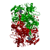
| |||||||||
| Unit cell |
| |||||||||
| Components on special symmetry positions |
|
-Components
-Protein , 1 types, 1 molecules A
| #1: Protein | Mass: 71763.766 Da / Num. of mol.: 1 / Fragment: AGAO HOLOENZYME, RESIDUES 3-638 Source method: isolated from a genetically manipulated source Details: RESIDUE A382 WAS AN ACTIVE SITE TYROSINE RESIDUE, WHICH WAS SELF-PROCESSED TO BECOME A TPQ Source: (gene. exp.) ARTHROBACTER GLOBIFORMIS (bacteria) / Plasmid: PAGAO2 / Production host: ESCHERICHIA COLI (E. coli) / Strain (production host): BL21(DE3) / References: UniProt: P46881, EC: 1.4.3.6 |
|---|
-Non-polymers , 6 types, 385 molecules 


| #2: Chemical | ChemComp-CU / Copper Mass: 63.546 Da / Num. of mol.: 1 / Source method: obtained synthetically / Formula: Cu Mass: 63.546 Da / Num. of mol.: 1 / Source method: obtained synthetically / Formula: Cu | ||||
|---|---|---|---|---|---|
| #3: Chemical | ChemComp-NA /  Mass: 22.990 Da / Num. of mol.: 1 / Source method: obtained synthetically / Formula: Na Mass: 22.990 Da / Num. of mol.: 1 / Source method: obtained synthetically / Formula: Na | ||||
| #4: Chemical | ChemComp-R6A /  Mass: 831.109 Da / Num. of mol.: 1 / Source method: obtained synthetically / Formula: C46H63N7ORu Mass: 831.109 Da / Num. of mol.: 1 / Source method: obtained synthetically / Formula: C46H63N7ORu | ||||
| #5: Chemical | Sulfate Mass: 96.063 Da / Num. of mol.: 3 / Source method: obtained synthetically / Formula: SO4 Mass: 96.063 Da / Num. of mol.: 3 / Source method: obtained synthetically / Formula: SO4#6: Chemical | ChemComp-GOL / Glycerol Mass: 92.094 Da / Num. of mol.: 5 / Source method: obtained synthetically / Formula: C3H8O3 Mass: 92.094 Da / Num. of mol.: 5 / Source method: obtained synthetically / Formula: C3H8O3#7: Water | ChemComp-HOH / | WaterMass: 18.015 Da / Num. of mol.: 374 / Source method: isolated from a natural source / Formula: H2O |
-Details
| Sequence details | RESIDES 1-2, MT, ARE THOUGHT TO BE CLEAVED. THE WILD TYPE MATURE PROTEIN IS COMPOSED OF RESIDUES 3- ...RESIDES 1-2, MT, ARE THOUGHT TO BE CLEAVED. THE WILD TYPE MATURE PROTEIN IS COMPOSED OF RESIDUES 3-638. RESIDUES 639- 640, SN, IS A PRE-TAG SPACER. RESIDUES 641-648, WSHPQFEK, IS A STREP-TAG II. SEE JUDA ET AL. (2001) PROTEIN EXPRESSION |
|---|
-Experimental details
-Experiment
| Experiment | Method: X-RAY DIFFRACTION / Number of used crystals: 1 |
|---|
- Sample preparation
| Crystal | Density Matthews: 3.07 Å3/Da / Density % sol: 59.99 % Description: STARTING MODEL IS A STRUCTURE NOT TO BE DEPOSITED THAT WAS IN TURN DERIVED FROM 1W6G |
|---|---|
| Crystal grow | Method: vapor diffusion, hanging drop / pH: 7 Details: THE CRYSTAL WAS GROWN BY HANGING DROP OVER 150 MM CITRATE PH 7.0, 700 MM NH4SO4. THE INHIBITOR WAS ADDED BY SOAKING, OVER SIX DAYS. 20 MG OF THE INHIBITOR (WC6) DISSOLVED IN 200 MICROLITRE ...Details: THE CRYSTAL WAS GROWN BY HANGING DROP OVER 150 MM CITRATE PH 7.0, 700 MM NH4SO4. THE INHIBITOR WAS ADDED BY SOAKING, OVER SIX DAYS. 20 MG OF THE INHIBITOR (WC6) DISSOLVED IN 200 MICROLITRE ETHANOL. 1MICROLITRE OF THIS WAS ADDED TO THE 200MICROLITRE RESERVOIR. GLYCEROL WAS THEN, ADDED IN 2-3% INCREMENTS, TO THE RESERVOIR, WITH THE NEW RESERVOIR SOLUTION SUBSTITUTING THE DROP SOLUTION. THE FINAL GLYCEROL CONCENTRATION WAS 30%. |
-Data collection
| Diffraction | Mean temperature: 100 K |
|---|---|
| Diffraction source | Source: ROTATING ANODE / Type: RIGAKU RU200 / Wavelength: 1.5418 |
| Detector | Type: MARRESEARCH / Detector: IMAGE PLATE / Date: Jan 23, 2004 / Details: OSMIC MIRRORS |
| Radiation | Protocol: SINGLE WAVELENGTH / Monochromatic (M) / Laue (L): M / Scattering type: x-ray |
| Radiation wavelength | Wavelength: 1.5418 Å / Relative weight: 1 |
| Reflection | Resolution: 1.79→15 Å / Num. obs: 474513 / % possible obs: 95 % / Observed criterion σ(I): -3 / Redundancy: 6.37 % / Biso Wilson estimate: 26.86 Å2 / Rmerge(I) obs: 0.04 / Net I/σ(I): 24.64 |
| Reflection shell | Resolution: 1.79→1.85 Å / Redundancy: 2.93 % / Rmerge(I) obs: 0.19 / Mean I/σ(I) obs: 3.44 / % possible all: 78.7 |
- Processing
| Software |
| ||||||||||||||||||||||||||||||||||||||||||||||||||||||||||||||||||||||||||||||||||||||||||||||||||||||||||||||||||||||||||||||||||||||||||||||||||||||||||||||||||||||||||||||||||||||
|---|---|---|---|---|---|---|---|---|---|---|---|---|---|---|---|---|---|---|---|---|---|---|---|---|---|---|---|---|---|---|---|---|---|---|---|---|---|---|---|---|---|---|---|---|---|---|---|---|---|---|---|---|---|---|---|---|---|---|---|---|---|---|---|---|---|---|---|---|---|---|---|---|---|---|---|---|---|---|---|---|---|---|---|---|---|---|---|---|---|---|---|---|---|---|---|---|---|---|---|---|---|---|---|---|---|---|---|---|---|---|---|---|---|---|---|---|---|---|---|---|---|---|---|---|---|---|---|---|---|---|---|---|---|---|---|---|---|---|---|---|---|---|---|---|---|---|---|---|---|---|---|---|---|---|---|---|---|---|---|---|---|---|---|---|---|---|---|---|---|---|---|---|---|---|---|---|---|---|---|---|---|---|---|
| Refinement | Method to determine structure: MOLECULAR REPLACEMENT / Resolution: 1.8→14.99 Å / Cor.coef. Fo:Fc: 0.973 / Cor.coef. Fo:Fc free: 0.966 / SU B: 2.346 / SU ML: 0.069 / Cross valid method: THROUGHOUT / ESU R: 0.095 / ESU R Free: 0.091 / Stereochemistry target values: MAXIMUM LIKELIHOOD / Details: HYDROGENS HAVE BEEN ADDED IN THE RIDING POSITIONS.
| ||||||||||||||||||||||||||||||||||||||||||||||||||||||||||||||||||||||||||||||||||||||||||||||||||||||||||||||||||||||||||||||||||||||||||||||||||||||||||||||||||||||||||||||||||||||
| Solvent computation | Ion probe radii: 0.8 Å / Shrinkage radii: 0.8 Å / VDW probe radii: 1.4 Å / Solvent model: BABINET MODEL WITH MASK | ||||||||||||||||||||||||||||||||||||||||||||||||||||||||||||||||||||||||||||||||||||||||||||||||||||||||||||||||||||||||||||||||||||||||||||||||||||||||||||||||||||||||||||||||||||||
| Displacement parameters | Biso mean: 29.12 Å2
| ||||||||||||||||||||||||||||||||||||||||||||||||||||||||||||||||||||||||||||||||||||||||||||||||||||||||||||||||||||||||||||||||||||||||||||||||||||||||||||||||||||||||||||||||||||||
| Refinement step | Cycle: LAST / Resolution: 1.8→14.99 Å
| ||||||||||||||||||||||||||||||||||||||||||||||||||||||||||||||||||||||||||||||||||||||||||||||||||||||||||||||||||||||||||||||||||||||||||||||||||||||||||||||||||||||||||||||||||||||
| Refine LS restraints |
|