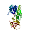
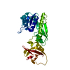
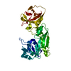
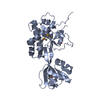
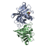
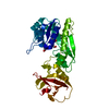
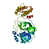
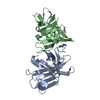
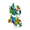
[English] 日本語
Yorodumi
- PDB-2cfi: The hydrolase domain of human 10-FTHFD in complex with 6- formylt... -

+
Open data


ID or keywords:

Loading...

-
Basic information

Entry
Database: PDB / ID: 2cfi
TitleThe hydrolase domain of human 10-FTHFD in complex with 6- formyltetrahydropterin
Components10-FORMYLTETRAHYDROFOLATE DEHYDROGENASE
KeywordsOXIDOREDUCTASE / TETRAHYDROFOLATE / FOLATE BINDING / NADP / ONE-CARBON METABOLISM / PHOSPHOPANTETHEINE
Function / homology
Function and homology information


formyltetrahydrofolate dehydrogenase / formyltetrahydrofolate dehydrogenase activity / 10-formyltetrahydrofolate catabolic process / NADPH regeneration / Metabolism of folate and pterines / aldehyde dehydrogenase (NAD+) activity / biosynthetic process / one-carbon metabolic process / extracellular exosome / cytosol
Similarity search - Function
Formyl transferase, C-terminal domain / Methionyl-tRNA Fmet Formyltransferase; Chain A, domain 2 / 10-formyltetrahydrofolate dehydrogenase / Formyl transferase, C-terminal domain superfamily / Formyl transferase, C-terminal / Formyl transferase, C-terminal domain / Formyl transferase-like, C-terminal domain superfamily / Formyl transferase, N-terminal domain / Phosphoribosylglycinamide formyltransferase, active site / Phosphoribosylglycinamide formyltransferase active site. ...Formyl transferase, C-terminal domain / Methionyl-tRNA Fmet Formyltransferase; Chain A, domain 2 / 10-formyltetrahydrofolate dehydrogenase / Formyl transferase, C-terminal domain superfamily / Formyl transferase, C-terminal / Formyl transferase, C-terminal domain / Formyl transferase-like, C-terminal domain superfamily / Formyl transferase, N-terminal domain / Phosphoribosylglycinamide formyltransferase, active site / Phosphoribosylglycinamide formyltransferase active site. / Formyl transferase, N-terminal / Formyl transferase / Formyl transferase, N-terminal domain superfamily / Aldehyde dehydrogenase, glutamic acid active site / Aldehyde dehydrogenases glutamic acid active site. / Aldehyde dehydrogenase, cysteine active site / Aldehyde dehydrogenases cysteine active site. / Aldehyde dehydrogenase domain / Aldehyde dehydrogenase family / Aldehyde dehydrogenase, N-terminal / Aldehyde dehydrogenase, C-terminal / Aldehyde/histidinol dehydrogenase / Phosphopantetheine attachment site / ACP-like superfamily / Carrier protein (CP) domain profile. / Phosphopantetheine binding ACP domain / Roll / Rossmann fold / 3-Layer(aba) Sandwich / Alpha Beta
Similarity search - Domain/homology
6-FORMYLTETRAHYDROPTERIN / Cytosolic 10-formyltetrahydrofolate dehydrogenase
Similarity search - Component
Biological speciesHOMO SAPIENS (human)
MethodX-RAY DIFFRACTION / SYNCHROTRON / MOLECULAR REPLACEMENT / Resolution: 1.85 Å
AuthorsKursula, P. / Stenmark, P. / Arrowsmith, C. / Edwards, A. / Ehn, M. / Graslund, S. / Hammarstrom, M. / Hallberg, M. / Kotenyova, T. / Nilsson-Ehle, P. ...Kursula, P. / Stenmark, P. / Arrowsmith, C. / Edwards, A. / Ehn, M. / Graslund, S. / Hammarstrom, M. / Hallberg, M. / Kotenyova, T. / Nilsson-Ehle, P. / Nordlund, P. / Ogg, D.J. / Persson, C. / Sagemark, J. / Schuler, H. / Sundstrom, M. / Thorsell, A. / Weigelt, J.
CitationJournal: Acta Crystallogr.,Sect.D / Year: 2006
Title: Structures of the Hydrolase Domain of Human 10-Formyltetrahydrofolate Dehydrogenase and its Complex with a Substrate Analogue.
Authors: Kursula, P. / Schuler, H. / Flodin, S. / Nilsson-Ehle, P. / Ogg, D.J. / Savitsky, P. / Nordlund, P. / Stenmark, P.
History
DepositionFeb 21, 2006Deposition site: PDBE / Processing site: PDBE
Revision 1.0Mar 14, 2006Provider: repository / Type: Initial release
Revision 1.1Jul 13, 2011Group: Advisory / Version format compliance
Revision 1.2Dec 13, 2023Group: Data collection / Database references ...Data collection / Database references / Other / Refinement description
Category: chem_comp_atom / chem_comp_bond ...chem_comp_atom / chem_comp_bond / database_2 / pdbx_database_status / pdbx_initial_refinement_model
Item: _database_2.pdbx_DOI / _database_2.pdbx_database_accession / _pdbx_database_status.status_code_sf
Remark 700 SHEET THE SHEET STRUCTURE OF THIS MOLECULE IS BIFURCATED. IN ORDER TO REPRESENT THIS FEATURE IN ... SHEET THE SHEET STRUCTURE OF THIS MOLECULE IS BIFURCATED. IN ORDER TO REPRESENT THIS FEATURE IN THE SHEET RECORDS BELOW, TWO SHEETS ARE DEFINED.

-
Structure visualization

Structure viewerMolecule:
MolmilJmol/JSmol

Downloads & links

-
Assembly

Deposited unit
A: 10-FORMYLTETRAHYDROFOLATE DEHYDROGENASE
hetero molecules


Theoretical massNumber of molelcules
Total (without water)36,8675
Polymers36,3831
Non-polymers4834
Water5,459303
1


  • Idetical with deposited unit
  • defined by software
TypeNameSymmetry operationNumber
identity operation1_555x,y,z1
MethodPISA
Unit cell
Length a, b, c (Å)108.140, 64.440, 58.870
Angle α, β, γ (deg.)90.00, 98.79, 90.00
Int Tables number5
Space group name H-MC121

-
Components

#1: Protein 10-FORMYLTETRAHYDROFOLATE DEHYDROGENASE / 10-FTHFDH / ALDEHYDE DEHYDROGENASE 1 FAMILY MEMBER L1


Mass: 36383.492 Da / Num. of mol.: 1 / Fragment: HYDROLASE DOMAIN, RESIDUES 1-307
Source method: isolated from a genetically manipulated source
Source: (gene. exp.) HOMO SAPIENS (human) / Plasmid: PNIC-BSA4 / Production host: ESCHERICHIA COLI (E. coli) / Strain (production host): BL21(DE3)
References: UniProt: O75891, formyltetrahydrofolate dehydrogenase
#2: Chemical ChemComp-SO4 / SULFATE ION / Sulfate


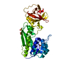
Mass: 96.063 Da / Num. of mol.: 3 / Source method: obtained synthetically / Formula: SO4
#3: Chemical ChemComp-ZZZ / 6-FORMYLTETRAHYDROPTERIN / (6S)-2-AMINO-4-OXO-3,4,5,6,7,8-HEXAHYDROPTERIDINE-6-CARBALDEHYDE


Mass: 195.179 Da / Num. of mol.: 1 / Source method: obtained synthetically / Formula: C7H9N5O2
#4: Water ChemComp-HOH / water / Water


Mass: 18.015 Da / Num. of mol.: 303 / Source method: isolated from a natural source / Formula: H2O

-
Experimental details

-
Experiment

ExperimentMethod: X-RAY DIFFRACTION / Number of used crystals: 1

-
Sample preparation

CrystalDensity Matthews: 2.61 Å3/Da / Density % sol: 52.52 %
Crystal growpH: 7.8 / Details: 1.4 M AMMONIUM SULFATE, 50 MM HEPES PH 7.8

-
Data collection

DiffractionMean temperature: 100 K
Diffraction sourceSource: SYNCHROTRON / Site: BESSY / Beamline: 14.1 / Wavelength: 0.9537
DetectorType: MARRESEARCH / Detector: CCD
RadiationProtocol: SINGLE WAVELENGTH / Monochromatic (M) / Laue (L): M / Scattering type: x-ray
Radiation wavelengthWavelength: 0.9537 Å / Relative weight: 1
ReflectionResolution: 1.85→20 Å / Num. obs: 34108 / % possible obs: 99.6 % / Observed criterion σ(I): -3 / Redundancy: 4.8 % / Rmerge(I) obs: 0.07 / Net I/σ(I): 14.3
Reflection shellResolution: 1.85→1.9 Å / Redundancy: 4.5 % / Rmerge(I) obs: 0.41 / Mean I/σ(I) obs: 4 / % possible all: 99.5

-
Processing

Software
NameVersionClassification
REFMAC5.2.0019refinement
XDSdata reduction
XSCALEdata scaling
RefinementMethod to determine structure: MOLECULAR REPLACEMENT
Starting model: PDB ENTRY 2BW0
Resolution: 1.85→10 Å / Cor.coef. Fo:Fc: 0.96 / Cor.coef. Fo:Fc free: 0.934 / SU B: 5.699 / SU ML: 0.089 / TLS residual ADP flag: LIKELY RESIDUAL / Cross valid method: THROUGHOUT / ESU R: 0.122 / ESU R Free: 0.124 / Stereochemistry target values: MAXIMUM LIKELIHOOD / Details: HYDROGENS HAVE BEEN ADDED IN THE RIDING POSITIONS.
RfactorNum. reflection% reflectionSelection details
Rfree0.225 1696 5 %RANDOM
Rwork0.178 ---
obs0.181 32226 99.7 %-
Solvent computationIon probe radii: 0.8 Å / Shrinkage radii: 0.8 Å / VDW probe radii: 1.4 Å / Solvent model: BABINET MODEL WITH MASK
Displacement parametersBiso mean: 31.77 Å2
Baniso -1Baniso -2Baniso -3
1-1.13 Å20 Å2-0.3 Å2
2---0.83 Å20 Å2
3----0.39 Å2
Refinement stepCycle: LAST / Resolution: 1.85→10 Å
ProteinNucleic acidLigandSolventTotal
Num. atoms2409 0 29 303 2741
Refine LS restraints
Refine-IDTypeDev idealDev ideal targetNumber
X-RAY DIFFRACTIONr_bond_refined_d0.0140.0222534
X-RAY DIFFRACTIONr_bond_other_d0.0020.021743
X-RAY DIFFRACTIONr_angle_refined_deg1.5751.9763443
X-RAY DIFFRACTIONr_angle_other_deg1.06834267
X-RAY DIFFRACTIONr_dihedral_angle_1_deg6.7665319
X-RAY DIFFRACTIONr_dihedral_angle_2_deg35.5924.595111
X-RAY DIFFRACTIONr_dihedral_angle_3_deg14.715430
X-RAY DIFFRACTIONr_dihedral_angle_4_deg17.0051512
X-RAY DIFFRACTIONr_chiral_restr0.160.2369
X-RAY DIFFRACTIONr_gen_planes_refined0.0050.022813
X-RAY DIFFRACTIONr_gen_planes_other0.0020.02499
X-RAY DIFFRACTIONr_nbd_refined0.2110.2530
X-RAY DIFFRACTIONr_nbd_other0.2010.21841
X-RAY DIFFRACTIONr_nbtor_refined0.1810.21206
X-RAY DIFFRACTIONr_nbtor_other0.0870.21288
X-RAY DIFFRACTIONr_xyhbond_nbd_refined0.1960.2205
X-RAY DIFFRACTIONr_xyhbond_nbd_other
X-RAY DIFFRACTIONr_metal_ion_refined
X-RAY DIFFRACTIONr_metal_ion_other
X-RAY DIFFRACTIONr_symmetry_vdw_refined0.1090.27
X-RAY DIFFRACTIONr_symmetry_vdw_other0.2180.235
X-RAY DIFFRACTIONr_symmetry_hbond_refined0.1810.224
X-RAY DIFFRACTIONr_symmetry_hbond_other
X-RAY DIFFRACTIONr_symmetry_metal_ion_refined
X-RAY DIFFRACTIONr_symmetry_metal_ion_other
X-RAY DIFFRACTIONr_mcbond_it1.34921608
X-RAY DIFFRACTIONr_mcbond_other
X-RAY DIFFRACTIONr_mcangle_it2.10432505
X-RAY DIFFRACTIONr_mcangle_other
X-RAY DIFFRACTIONr_scbond_it2.5441075
X-RAY DIFFRACTIONr_scbond_other
X-RAY DIFFRACTIONr_scangle_it3.5015933
X-RAY DIFFRACTIONr_scangle_other
X-RAY DIFFRACTIONr_long_range_B_refined
X-RAY DIFFRACTIONr_long_range_B_other
X-RAY DIFFRACTIONr_rigid_bond_restr
X-RAY DIFFRACTIONr_sphericity_free
X-RAY DIFFRACTIONr_sphericity_bonded
LS refinement shellResolution: 1.85→1.9 Å / Total num. of bins used: 20 /
RfactorNum. reflection
Rfree0.29 122
Rwork0.225 2306
Refinement TLS params.Method: refined / Origin x: 27.7766 Å / Origin y: -0.037 Å / Origin z: 16.8999 Å
111213212223313233
T-0.2017 Å20.0246 Å2-0.0034 Å2--0.1771 Å20.0163 Å2---0.1371 Å2
L0.9526 °20.6581 °20.5028 °2-1.882 °21.7058 °2--2.9007 °2
S-0.0324 Å °-0.0474 Å °0.1772 Å °-0.1306 Å °-0.1159 Å °0.2372 Å °-0.1385 Å °-0.1742 Å °0.1483 Å °
Refinement TLS group
IDRefine-IDRefine TLS-IDAuth asym-IDAuth seq-ID
1X-RAY DIFFRACTION1A-2 - 307
2X-RAY DIFFRACTION1A1311

+
About Yorodumi

-
News

-
Feb 9, 2022. New format data for meta-information of EMDB entries

New format data for meta-information of EMDB entries

  • Version 3 of the EMDB header file is now the official format.
  • The previous official version 1.9 will be removed from the archive.

Related info.:EMDB header

External links:wwPDB to switch to version 3 of the EMDB data model

-
Aug 12, 2020. Covid-19 info

Covid-19 info

URL: https://pdbj.org/emnavi/covid19.php

New page: Covid-19 featured information page in EM Navigator.

Related info.:Covid-19 info / Mar 5, 2020. Novel coronavirus structure data

+
Mar 5, 2020. Novel coronavirus structure data

Novel coronavirus structure data

Related info.:Yorodumi Speices / Aug 12, 2020. Covid-19 info

External links:COVID-19 featured content - PDBj / Molecule of the Month (242):Coronavirus Proteases

+
Jan 31, 2019. EMDB accession codes are about to change! (news from PDBe EMDB page)

EMDB accession codes are about to change! (news from PDBe EMDB page)

  • The allocation of 4 digits for EMDB accession codes will soon come to an end. Whilst these codes will remain in use, new EMDB accession codes will include an additional digit and will expand incrementally as the available range of codes is exhausted. The current 4-digit format prefixed with “EMD-” (i.e. EMD-XXXX) will advance to a 5-digit format (i.e. EMD-XXXXX), and so on. It is currently estimated that the 4-digit codes will be depleted around Spring 2019, at which point the 5-digit format will come into force.
  • The EM Navigator/Yorodumi systems omit the EMD- prefix.

Related info.:Q: What is EMD? / ID/Accession-code notation in Yorodumi/EM Navigator

External links:EMDB Accession Codes are Changing Soon! / Contact to PDBj

+
Jul 12, 2017. Major update of PDB

Major update of PDB

  • wwPDB released updated PDB data conforming to the new PDBx/mmCIF dictionary.
  • This is a major update changing the version number from 4 to 5, and with Remediation, in which all the entries are updated.
  • In this update, many items about electron microscopy experimental information are reorganized (e.g. em_software).
  • Now, EM Navigator and Yorodumi are based on the updated data.

External links:wwPDB Remediation / Enriched Model Files Conforming to OneDep Data Standards Now Available in the PDB FTP Archive

-
Yorodumi

Thousand views of thousand structures

  • Yorodumi is a browser for structure data from EMDB, PDB, SASBDB, etc.
  • This page is also the successor to EM Navigator detail page, and also detail information page/front-end page for Omokage search.
  • The word "yorodu" (or yorozu) is an old Japanese word meaning "ten thousand". "mi" (miru) is to see.

Related info.:EMDB / PDB / SASBDB / Comparison of 3 databanks / Yorodumi Search / Aug 31, 2016. New EM Navigator & Yorodumi / Yorodumi Papers / Jmol/JSmol / Function and homology information / Changes in new EM Navigator and Yorodumi

Read more