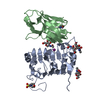
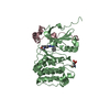
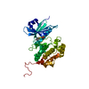
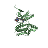
Entry Database : PDB / ID : 2bujTitle Crystal structure of the human Serine-threonine Kinase 16 in complex with staurosporine SERINE/THREONINE-PROTEIN KINASE 16 Keywords / / / / / / / Function / homology Function Domain/homology Component
/ / / / / / / / / / / / / / / / / / / / / / / / / / / / / / / / / / / / / / / / / / / / / / / / / / Biological species HOMO SAPIENS (human)Method / / / Resolution : 2.6 Å Authors Debreczeni, J.E. / Eswaran, J. / Bullock, A. / Filippakopoulos, P. / Kavanagh, K. / Amos, A. / Fedorov, O. / Sobott, F. / Ball, L.J. / von Delft, F. ...Debreczeni, J.E. / Eswaran, J. / Bullock, A. / Filippakopoulos, P. / Kavanagh, K. / Amos, A. / Fedorov, O. / Sobott, F. / Ball, L.J. / von Delft, F. / Arrowsmith, C. / Sundstrom, M. / Edwards, A. / Knapp, S. Journal : To be Published Title : Crystal Structure of the Human Serine-Threonine Kinase 16 in Complex with StaurosporineAuthors : Debreczeni, J.E. / Eswaran, J. / Bullock, A. / Filippakopoulos, P. / Kavanagh, K. / Amos, A. / Fedorov, O. / Sobott, F. / Ball, L.J. / von Delft, F. / Arrowsmith, C. / Sundstrom, M. / Edwards, A. / Knapp, S. History Deposition Jun 13, 2005 Deposition site / Processing site Revision 1.0 Jul 5, 2005 Provider / Type Revision 1.1 Jul 13, 2011 Group / Refinement description / Version format complianceRevision 1.2 Jan 24, 2018 Group / Structure summary / Category / citation_author / Item / _citation_author.nameRevision 1.3 Apr 10, 2019 Group / Source and taxonomy / Category / struct_biolItem / _entity_src_gen.pdbx_host_org_variantRevision 1.4 May 8, 2019 Group / Experimental preparation / Category / Item Revision 1.5 May 8, 2024 Group / Database references / OtherCategory chem_comp_atom / chem_comp_bond ... chem_comp_atom / chem_comp_bond / database_2 / pdbx_database_status Item / _database_2.pdbx_database_accession / _pdbx_database_status.status_code_sf
Show all Show less
 Movie
Movie Controller

 Yorodumi
 Open data
 Basic information
 Components
 Keywords
 Function and homology information
 HOMO SAPIENS (human)
 X-RAY DIFFRACTION /
  Authors
 Citation
 Structure visualization
 Downloads & links
 Other downloads
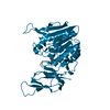


 PDBj
PDBj Assembly
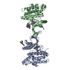


 Mass: 35.453 Da / Num. of mol.: 2 / Source method: obtained synthetically / Formula: Cl
 Mass: 466.531 Da / Num. of mol.: 4 / Source method: obtained synthetically / Formula: C28H26N4O3 / Comment: anticancer, antifungal, antibiotic, alkaloid*YM
 Mass: 18.015 Da / Num. of mol.: 76 / Source method: isolated from a natural source / Formula: H2O
 Sample preparation
 / Beamline: X10SA / Wavelength: 0.984
  Processing