 Movie
Movie Controller

[English] 日本語
 Yorodumi
- PDB-2bsx: Crystal structure of the Plasmodium falciparum purine nucleoside ... -
+ Open data

- Basic information
| Entry | Database: PDB / ID: 2bsx | ||||||
|---|---|---|---|---|---|---|---|
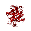
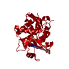
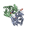
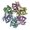
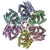
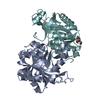
| Title | Crystal structure of the Plasmodium falciparum purine nucleoside phosphorylase complexed with inosine | ||||||
 Components Components | PURINE NUCLEOSIDE PHOSPHORYLASE | ||||||
 Keywords Keywords | TRANSFERASE / PURINE NUCLEOSIDE PHOSPHORYLASE / URIDINE PHOSPHORYLASE / PUTATIVE / GLYCOSYLTRANSFERASE | ||||||
| Function / homology |  Function and homology information Function and homology informationPyrimidine salvage / Pyrimidine catabolism / S-methyl-5'-thioinosine phosphorylase / purine nucleotide catabolic process / uridine catabolic process / inosine catabolic process / S-methyl-5-thioadenosine phosphorylase activity / guanosine phosphorylase activity / uridine phosphorylase activity / purine-nucleoside phosphorylase ...Pyrimidine salvage / Pyrimidine catabolism / S-methyl-5'-thioinosine phosphorylase / purine nucleotide catabolic process / uridine catabolic process / inosine catabolic process / S-methyl-5-thioadenosine phosphorylase activity / guanosine phosphorylase activity / uridine phosphorylase activity / purine-nucleoside phosphorylase / purine-nucleoside phosphorylase activity / purine ribonucleoside salvage / cytosol Similarity search - Function | ||||||
| Biological species |  | ||||||
| Method |  X-RAY DIFFRACTION / SYNCHROTRON / MOLECULAR REPLACEMENT / Resolution: 2 Å X-RAY DIFFRACTION / SYNCHROTRON / MOLECULAR REPLACEMENT / Resolution: 2 Å | ||||||
 Authors Authors | Schnick, C. / Brzozowski, A.M. / Dodson, E.J. / Murshudov, G.N. / Brannigan, J.A. / Wilkinson, A.J. | ||||||
 Citation Citation | Journal: Acta Crystallogr.,Sect.D / Year: 2005 Title: Structures of Plasmodium Falciparum Purine Nucleoside Phosphorylase Complexed with Sulfate and its Natural Substrate Inosine Authors: Schnick, C. / Robien, M.A. / Brzozowski, A.M. / Dodson, E.J. / Murshudov, G.N. / Anderson, L. / Luft, J.R. / Mehlin, C. / Hol, W.G.J. / Brannigan, J.A. / Wilkinson, A.J. | ||||||
| History |
| ||||||
| Remark 700 | SHEET THE SHEET STRUCTURE OF THIS MOLECULE IS BIFURCATED. IN ORDER TO REPRESENT THIS FEATURE IN ... SHEET THE SHEET STRUCTURE OF THIS MOLECULE IS BIFURCATED. IN ORDER TO REPRESENT THIS FEATURE IN THE SHEET RECORDS BELOW, TWO SHEETS ARE DEFINED. |
- Structure visualization
| Structure viewer | Molecule: MolmilJmol/JSmol |
|---|
- Downloads & links
-Download
| PDBx/mmCIF format | 2bsx.cif.gz | 62.7 KB | Display | PDBx/mmCIF format |
|---|---|---|---|---|
| PDB format | pdb2bsx.ent.gz | 45.5 KB | Display | PDB format |
| PDBx/mmJSON format | 2bsx.json.gz | Tree view | PDBx/mmJSON format | |
| Others |  Other downloads Other downloads |
-Validation report
| Arichive directory | https://data.pdbj.org/pub/pdb/validation_reports/bs/2bsxftp://data.pdbj.org/pub/pdb/validation_reports/bs/2bsx | HTTPS FTP |
|---|
-Related structure data
| Related structure data |  1sq6C  1lx7S S: Starting model for refinement C: citing same article ( |
|---|---|
| Similar structure data |


-Links
 PDBj
PDBj- Assembly
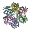
| Deposited unit | 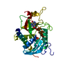
| |||||||||
|---|---|---|---|---|---|---|---|---|---|---|
| 1 | x 6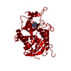
| |||||||||
| Unit cell |
| |||||||||
| Components on special symmetry positions |
|
-Components
| #1: Protein | Mass: 27962.311 Da / Num. of mol.: 1 Source method: isolated from a genetically manipulated source Source: (gene. exp.) Strain: 3D7 / Plasmid: PET28A / Production host:  References: UniProt: Q8T9Z7, UniProt: Q8I3X4*PLUS, purine-nucleoside phosphorylase |
|---|---|
| #2: Chemical | ChemComp-NOS /   Mass: 268.226 Da / Num. of mol.: 1 / Source method: obtained synthetically / Formula: C10H12N4O5 Mass: 268.226 Da / Num. of mol.: 1 / Source method: obtained synthetically / Formula: C10H12N4O5 |
| #3: Water | ChemComp-HOH /  Mass: 18.015 Da / Num. of mol.: 105 / Source method: isolated from a natural source / Formula: H2O Mass: 18.015 Da / Num. of mol.: 105 / Source method: isolated from a natural source / Formula: H2O |
| Has protein modification | Y |
| Sequence details | RESIDUES 246-253 INSERTION FOR CLONING PURPOSE |
-Experimental details
-Experiment
| Experiment | Method: X-RAY DIFFRACTION |
|---|
- Sample preparation
| Crystal | Density Matthews: 2.3 Å3/Da / Density % sol: 45.6 % |
|---|---|
| Crystal grow | pH: 7.5 Details: 10 % PEG 8000, 0.1 M HEPES PH 7.5, 30 % HEXANETRIOL |
-Data collection
| Diffraction | Mean temperature: 100 K |
|---|---|
| Diffraction source | Source: SYNCHROTRON / Site: ESRF  / Beamline: ID29 / Wavelength: 0.9168 / Beamline: ID29 / Wavelength: 0.9168 |
| Detector | Type: ADSC CCD / Detector: CCD / Date: Sep 12, 2003 |
| Radiation | Protocol: SINGLE WAVELENGTH / Monochromatic (M) / Laue (L): M / Scattering type: x-ray |
| Radiation wavelength | Wavelength: 0.9168 Å / Relative weight: 1 |
| Reflection | Resolution: 2→20 Å / Num. obs: 27984 / % possible obs: 87 % / Observed criterion σ(I): 0 / Redundancy: 2.5 % / Rmerge(I) obs: 0.08 / Net I/σ(I): 10 |
| Reflection shell | Resolution: 2→2.07 Å / Redundancy: 2.4 % / Rmerge(I) obs: 0.35 / Mean I/σ(I) obs: 5 / % possible all: 80 |
- Processing
| Software |
| ||||||||||||||||||||||||||||||||||||||||||||||||||||||||||||||||||||||||||||||||||||||||||||||||||||||||||||||||||||||||||||||||||||||||||||||||||||||||||||||||||||||||||||||||||||||
|---|---|---|---|---|---|---|---|---|---|---|---|---|---|---|---|---|---|---|---|---|---|---|---|---|---|---|---|---|---|---|---|---|---|---|---|---|---|---|---|---|---|---|---|---|---|---|---|---|---|---|---|---|---|---|---|---|---|---|---|---|---|---|---|---|---|---|---|---|---|---|---|---|---|---|---|---|---|---|---|---|---|---|---|---|---|---|---|---|---|---|---|---|---|---|---|---|---|---|---|---|---|---|---|---|---|---|---|---|---|---|---|---|---|---|---|---|---|---|---|---|---|---|---|---|---|---|---|---|---|---|---|---|---|---|---|---|---|---|---|---|---|---|---|---|---|---|---|---|---|---|---|---|---|---|---|---|---|---|---|---|---|---|---|---|---|---|---|---|---|---|---|---|---|---|---|---|---|---|---|---|---|---|---|
| Refinement | Method to determine structure: MOLECULAR REPLACEMENT Starting model: PDB ENTRY 1LX7 Resolution: 2→18.9 Å / Cor.coef. Fo:Fc: 0.921 / Cor.coef. Fo:Fc free: 0.852 / SU B: 8.454 / SU ML: 0.237 / Cross valid method: THROUGHOUT / ESU R: 0.299 / ESU R Free: 0.262 / Stereochemistry target values: MAXIMUM LIKELIHOOD Details: HYDROGENS HAVE BEEN ADDED IN THE RIDING POSITIONS. RESIDUES 215-220 ARE DISORDERED.
| ||||||||||||||||||||||||||||||||||||||||||||||||||||||||||||||||||||||||||||||||||||||||||||||||||||||||||||||||||||||||||||||||||||||||||||||||||||||||||||||||||||||||||||||||||||||
| Solvent computation | Ion probe radii: 0.8 Å / Shrinkage radii: 0.8 Å / VDW probe radii: 1.2 Å / Solvent model: MASK | ||||||||||||||||||||||||||||||||||||||||||||||||||||||||||||||||||||||||||||||||||||||||||||||||||||||||||||||||||||||||||||||||||||||||||||||||||||||||||||||||||||||||||||||||||||||
| Displacement parameters | Biso mean: 38.57 Å2
| ||||||||||||||||||||||||||||||||||||||||||||||||||||||||||||||||||||||||||||||||||||||||||||||||||||||||||||||||||||||||||||||||||||||||||||||||||||||||||||||||||||||||||||||||||||||
| Refinement step | Cycle: LAST / Resolution: 2→18.9 Å
| ||||||||||||||||||||||||||||||||||||||||||||||||||||||||||||||||||||||||||||||||||||||||||||||||||||||||||||||||||||||||||||||||||||||||||||||||||||||||||||||||||||||||||||||||||||||
| Refine LS restraints |
|