 Movie
Movie Controller

[English] 日本語
 Yorodumi
- PDB-2bp2: THE STRUCTURE OF BOVINE PANCREATIC PROPHOSPHOLIPASE A2 AT 3.0 ANG... -
+ Open data

- Basic information
| Entry | Database: PDB / ID: 2bp2 | ||||||
|---|---|---|---|---|---|---|---|
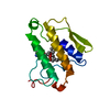
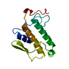
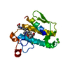
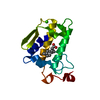
| Title | THE STRUCTURE OF BOVINE PANCREATIC PROPHOSPHOLIPASE A2 AT 3.0 ANGSTROMS RESOLUTION | ||||||
 Components Components | PHOSPHOLIPASE A2 | ||||||
 Keywords Keywords | HYDROLASE ZYMOGEN | ||||||
| Function / homology |  Function and homology information Function and homology informationAcyl chain remodelling of PS / Acyl chain remodelling of PG / Synthesis of PA / Acyl chain remodelling of PC / Acyl chain remodelling of PE / Acyl chain remodelling of PI / positive regulation of podocyte apoptotic process / phosphatidylglycerol metabolic process / phosphatidylcholine metabolic process / bile acid binding ...Acyl chain remodelling of PS / Acyl chain remodelling of PG / Synthesis of PA / Acyl chain remodelling of PC / Acyl chain remodelling of PE / Acyl chain remodelling of PI / positive regulation of podocyte apoptotic process / phosphatidylglycerol metabolic process / phosphatidylcholine metabolic process / bile acid binding / phospholipase A2 / : / arachidonate secretion / lipid catabolic process / innate immune response in mucosa / phospholipid binding / positive regulation of fibroblast proliferation / fatty acid biosynthetic process / antimicrobial humoral immune response mediated by antimicrobial peptide / antibacterial humoral response / defense response to Gram-positive bacterium / signaling receptor binding / calcium ion binding / cell surface / extracellular space Similarity search - Function | ||||||
| Biological species |  | ||||||
| Method |  X-RAY DIFFRACTION / Resolution: 3 Å X-RAY DIFFRACTION / Resolution: 3 Å | ||||||
 Authors Authors | Dijkstra, B.W. / Vannes, G.J.H. / Kalk, K.H. / Brandenburg, N.P. / Hol, W.G.J. / Drenth, J. | ||||||
 Citation Citation | Journal: Acta Crystallogr.,Sect.B / Year: 1982 Title: The Structure of Bovine Pancreatic Prophospholipase A2 at 3.0 Angstroms Resolution Authors: Dijkstra, B.W. / Vannes, G.J.H. / Kalk, K.H. / Brandenburg, N.P. / Hol, W.G.J. / Drenth, J. #1: Journal: J.Mol.Biol. / Year: 1981Title: Structure of Bovine Pancreatic Phospholipase A2 at 1.7 Angstroms Resolution Authors: Dijkstra, B.W. / Kalk, K.H. / Hol, W.G.J. / Drenth, J. #2: Journal: Nature / Year: 1981Title: Active Site and Catalytic Mechanism of Phospholipase A2 Authors: Dijkstra, B.W. / Drenth, J. / Kalk, K.H. #3: Journal: Thesis / Year: 1980Title: Structure and Mechanism of Phospholipase A2 Authors: Dijkstra, B.W. #4: Journal: J.Mol.Biol. / Year: 1978Title: Three-Dimensional Structure and Disulfide Bond Connections in Bovine Pancreatic Phospholipase A2 Authors: Dijkstra, B.W. / Drenth, J. / Kalk, K.H. / Vandermaelen, P. | ||||||
| History |
|
- Structure visualization
| Structure viewer | Molecule: MolmilJmol/JSmol |
|---|
- Downloads & links
-Download
| PDBx/mmCIF format | 2bp2.cif.gz | 35.6 KB | Display | PDBx/mmCIF format |
|---|---|---|---|---|
| PDB format | pdb2bp2.ent.gz | 24.7 KB | Display | PDB format |
| PDBx/mmJSON format | 2bp2.json.gz | Tree view | PDBx/mmJSON format | |
| Others |  Other downloads Other downloads |
-Validation report
| Arichive directory | https://data.pdbj.org/pub/pdb/validation_reports/bp/2bp2ftp://data.pdbj.org/pub/pdb/validation_reports/bp/2bp2 | HTTPS FTP |
|---|
-Related structure data
| Similar structure data |
|---|


-Links
 PDBj
PDBj

- Assembly
| Deposited unit | 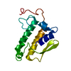
| ||||||||
|---|---|---|---|---|---|---|---|---|---|
| 1 |
| ||||||||
| Unit cell |
| ||||||||
| Atom site foot note | 1: SEE REMARK 8 ABOVE. |
-Components
| #1: Protein | Mass: 14539.279 Da / Num. of mol.: 1 Source method: isolated from a genetically manipulated source Source: (gene. exp.) |
|---|---|
| Compound details | THE ZYMOGEN PROPHOSPHOLIPASE A2 CONTAINS SEVEN EXTRA RESIDUES AT THE N-TERMINUS COMPARED WITH THE ...THE ZYMOGEN PROPHOSPHO |
| Has protein modification | Y |
| Sequence details | SEQUENCE NUMBERING IS THE SAME AS FOR THE ACTIVE ENZYME, I. E. THE FIRST RESIDUE IS NUMBERED -7. |
-Experimental details
-Experiment
| Experiment | Method: X-RAY DIFFRACTION |
|---|
- Sample preparation
| Crystal | Density Matthews: 2.23 Å3/Da / Density % sol: 44.9 % | |||||||||||||||||||||||||||||||||||
|---|---|---|---|---|---|---|---|---|---|---|---|---|---|---|---|---|---|---|---|---|---|---|---|---|---|---|---|---|---|---|---|---|---|---|---|---|
| Crystal grow | *PLUS pH: 7.6 / Method: unknown / Details: Dijkstra, B.W., (1978) J.Mol.Biol., 124, 53. | |||||||||||||||||||||||||||||||||||
| Components of the solutions | *PLUS
|
- Processing
| Software | Name: PROLSQ / Classification: refinement | ||||||||||||
|---|---|---|---|---|---|---|---|---|---|---|---|---|---|
| Refinement | Rfactor Rwork: 0.219 / Highest resolution: 3 Å Details: RESIDUES 1 TO 3 INCLUSIVE AND 62 TO 73 INCLUSIVE ARE VIRTUALLY INVISIBLE IN ELECTRON DENSITY MAPS AND ARE PROBABLY DISORDERED. THE COORDINATES GIVEN BELOW FOR THESE RESIDUES CONTAIN, ...Details: RESIDUES 1 TO 3 INCLUSIVE AND 62 TO 73 INCLUSIVE ARE VIRTUALLY INVISIBLE IN ELECTRON DENSITY MAPS AND ARE PROBABLY DISORDERED. THE COORDINATES GIVEN BELOW FOR THESE RESIDUES CONTAIN, THEREFORE, VERY LARGE ERRORS. | ||||||||||||
| Refinement step | Cycle: LAST / Highest resolution: 3 Å
| ||||||||||||
| Refinement | *PLUS Lowest resolution: 7.1 Å / Rfactor obs: 0.219 | ||||||||||||
| Solvent computation | *PLUS | ||||||||||||
| Displacement parameters | *PLUS |
