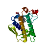
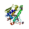
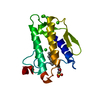
- PDB-2b04: Crystal Structure of Porcine Pancreatic Phospholipase A2 in Compl... -
+
Open data
ID or keywords:
Loading...
-
Basic information
Entry
Database: PDB / ID: 2b04
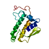
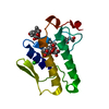
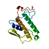
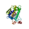
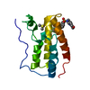
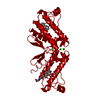
Title
Crystal Structure of Porcine Pancreatic Phospholipase A2 in Complex with Glycochenodeoxycholate
Components
Phospholipase A2, major isoenzyme
Keywords
HYDROLASE / Bile salt / glycochenodeoxycholate / carboxylic ester hydrolase / pancreatic enzyme
Function / homology
Function and homology information
regulation of D-glucose import across plasma membrane / positive regulation of podocyte apoptotic process / phosphatidylglycerol metabolic process / phosphatidylcholine metabolic process / A2-type glycerophospholipase activity / leukotriene biosynthetic process / : / bile acid binding / phospholipase A2 / neutrophil mediated immunity ...regulation of D-glucose import across plasma membrane / positive regulation of podocyte apoptotic process / phosphatidylglycerol metabolic process / phosphatidylcholine metabolic process / A2-type glycerophospholipase activity / leukotriene biosynthetic process / : / bile acid binding / phospholipase A2 / neutrophil mediated immunity / positive regulation of calcium ion transport into cytosol / lipid catabolic process / neutrophil chemotaxis / positive regulation of interleukin-8 production / phospholipid binding / positive regulation of immune response / positive regulation of fibroblast proliferation / fatty acid biosynthetic process / cellular response to insulin stimulus / positive regulation of MAPK cascade / intracellular signal transduction / signaling receptor binding / calcium ion binding / positive regulation of cell population proliferation / cell surface / positive regulation of transcription by RNA polymerase II / extracellular region Similarity search - Function
Phospholipase A2, aspartic acid active site / Phospholipase A2 aspartic acid active site. / Phospholipase A2, histidine active site / Phospholipase A2 histidine active site. / Phospholipase A2 / Phospholipase A2 domain / Phospholipase A2 / Phospholipase A2 / Phospholipase A2 domain / Phospholipase A2 ...Phospholipase A2, aspartic acid active site / Phospholipase A2 aspartic acid active site. / Phospholipase A2, histidine active site / Phospholipase A2 histidine active site. / Phospholipase A2 / Phospholipase A2 domain / Phospholipase A2 / Phospholipase A2 / Phospholipase A2 domain / Phospholipase A2 / Phospholipase A2 domain superfamily / Up-down Bundle / Mainly Alpha Similarity search - Domain/homology
Resolution: 2→2.07 Å / Redundancy: 5 % / Rmerge(I) obs: 0.421 / Mean I/σ(I) obs: 2.4 / Rsym value: 0.259 / % possible all: 99.7
-
Processing
Software
Name
Version
Classification
CNS
1.1
refinement
DENZO
datareduction
SCALEPACK
datascaling
AMoRE
phasing
Refinement
Method to determine structure: MOLECULAR REPLACEMENT / Resolution: 2.5→24.07 Å / Rfactor Rfree error: 0.014 / Data cutoff high absF: 888178.04 / Data cutoff low absF: 0 / Isotropic thermal model: RESTRAINED / Cross valid method: THROUGHOUT / σ(F): 3 / Stereochemistry target values: Engh & Huber Details: Residues A16 to A21 were deleted from the model due to disorder.
In the structure databanks used in Yorodumi, some data are registered as the other names, "COVID-19 virus" and "2019-nCoV". Here are the details of the virus and the list of structure data.
Jan 31, 2019. EMDB accession codes are about to change! (news from PDBe EMDB page)
EMDB accession codes are about to change! (news from PDBe EMDB page)
The allocation of 4 digits for EMDB accession codes will soon come to an end. Whilst these codes will remain in use, new EMDB accession codes will include an additional digit and will expand incrementally as the available range of codes is exhausted. The current 4-digit format prefixed with “EMD-” (i.e. EMD-XXXX) will advance to a 5-digit format (i.e. EMD-XXXXX), and so on. It is currently estimated that the 4-digit codes will be depleted around Spring 2019, at which point the 5-digit format will come into force.
The EM Navigator/Yorodumi systems omit the EMD- prefix.
Related info.:Q: What is EMD? / ID/Accession-code notation in Yorodumi/EM Navigator
Yorodumi is a browser for structure data from EMDB, PDB, SASBDB, etc.
This page is also the successor to EM Navigator detail page, and also detail information page/front-end page for Omokage search.
The word "yorodu" (or yorozu) is an old Japanese word meaning "ten thousand". "mi" (miru) is to see.
Related info.:EMDB / PDB / SASBDB / Comparison of 3 databanks / Yorodumi Search / Aug 31, 2016. New EM Navigator & Yorodumi / Yorodumi Papers / Jmol/JSmol / Function and homology information / Changes in new EM Navigator and Yorodumi
 Movie
Movie Controller

 Yorodumi
 Open data
 Basic information
 Components
 Keywords
 Function and homology information
 X-RAY DIFFRACTION /
  Authors
 Citation
 Structure visualization
 Downloads & links
 Other downloads
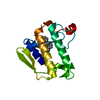


 PDBj
PDBj

 Assembly
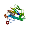

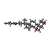
 Mass: 449.623 Da / Num. of mol.: 1
 Mass: 40.078 Da / Num. of mol.: 3 / Source method: obtained synthetically / Formula: Ca
 Mass: 35.453 Da / Num. of mol.: 1 / Source method: obtained synthetically / Formula: Cl
 Mass: 18.015 Da / Num. of mol.: 96 / Source method: isolated from a natural source / Formula: H2O
 Sample preparation
 Processing