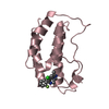
Group: Atomic model / Data collection ...Atomic model / Data collection / Database references / Derived calculations / Refinement description / Source and taxonomy / Structure summary Category: atom_site / citation ...atom_site / citation / citation_author / database_2 / entity_src_gen / pdbx_contact_author / pdbx_nonpoly_scheme / refine / refine_hist / refine_ls_shell / reflns / reflns_shell / software / struct_site / struct_site_gen Item: _atom_site.Cartn_x / _atom_site.Cartn_y ..._atom_site.Cartn_x / _atom_site.Cartn_y / _atom_site.Cartn_z / _citation.country / _citation.journal_abbrev / _citation.journal_id_ASTM / _citation.journal_id_CSD / _citation.journal_id_ISSN / _citation.pdbx_database_id_DOI / _citation.title / _citation.year / _database_2.pdbx_DOI / _database_2.pdbx_database_accession / _entity_src_gen.gene_src_common_name / _pdbx_contact_author.id / _pdbx_nonpoly_scheme.auth_mon_id / _pdbx_nonpoly_scheme.auth_seq_num / _refine.B_iso_mean / _refine.ls_R_factor_R_free / _refine.ls_R_factor_R_work / _refine.ls_R_factor_obs / _refine.ls_d_res_high / _refine.ls_d_res_low / _refine.ls_number_reflns_R_work / _refine_hist.d_res_high / _refine_hist.d_res_low / _refine_ls_shell.d_res_high / _refine_ls_shell.d_res_low / _reflns.pdbx_number_measured_all / _reflns.pdbx_scaling_rejects / _software.classification / _software.name / _software.version Description: Ligand geometry Details: The inhibitor, ZL0590, was associated with the binding site of a symmetry related protomer in the crystal lattice. This structure changes the coordinates for the ZL0590 inhibitor so that it ...Details: The inhibitor, ZL0590, was associated with the binding site of a symmetry related protomer in the crystal lattice. This structure changes the coordinates for the ZL0590 inhibitor so that it is in the binding site that agrees with the structure activity relationship for this compound series. Provider: author / Type: Coordinate replacement
In the structure databanks used in Yorodumi, some data are registered as the other names, "COVID-19 virus" and "2019-nCoV". Here are the details of the virus and the list of structure data.
Jan 31, 2019. EMDB accession codes are about to change! (news from PDBe EMDB page)
EMDB accession codes are about to change! (news from PDBe EMDB page)
The allocation of 4 digits for EMDB accession codes will soon come to an end. Whilst these codes will remain in use, new EMDB accession codes will include an additional digit and will expand incrementally as the available range of codes is exhausted. The current 4-digit format prefixed with “EMD-” (i.e. EMD-XXXX) will advance to a 5-digit format (i.e. EMD-XXXXX), and so on. It is currently estimated that the 4-digit codes will be depleted around Spring 2019, at which point the 5-digit format will come into force.
The EM Navigator/Yorodumi systems omit the EMD- prefix.
Related info.:Q: What is EMD? / ID/Accession-code notation in Yorodumi/EM Navigator
Yorodumi is a browser for structure data from EMDB, PDB, SASBDB, etc.
This page is also the successor to EM Navigator detail page, and also detail information page/front-end page for Omokage search.
The word "yorodu" (or yorozu) is an old Japanese word meaning "ten thousand". "mi" (miru) is to see.
Related info.:EMDB / PDB / SASBDB / Comparison of 3 databanks / Yorodumi Search / Aug 31, 2016. New EM Navigator & Yorodumi / Yorodumi Papers / Jmol/JSmol / Function and homology information / Changes in new EM Navigator and Yorodumi
 Movie
Movie Controller

 Yorodumi
 Open data
 Basic information
 Components
 Keywords
 Function and homology information
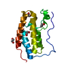
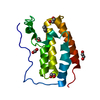
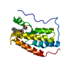
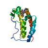
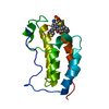
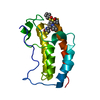
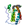
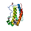
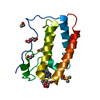
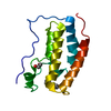
 Homo sapiens (human)
 X-RAY DIFFRACTION /
  Authors
 United States, 1items
  Citation
 Structure visualization
 Downloads & links
 Other downloads


 PDBj
PDBj Assembly
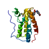


 Mass: 78.133 Da / Num. of mol.: 1 / Source method: obtained synthetically / Formula: C2H6OS / Comment: DMSO, precipitant*YM
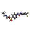
 Mass: 512.545 Da / Num. of mol.: 1 / Source method: obtained synthetically / Formula: C23H27F3N4O4S / Feature type: SUBJECT OF INVESTIGATION
 Mass: 18.015 Da / Num. of mol.: 185 / Source method: isolated from a natural source / Formula: H2O
 Sample preparation
 Processing