 Movie
Movie Controller

[English] 日本語
 Yorodumi
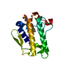
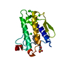
- PDB-2azy: Crystal Structure of Porcine Pancreatic Phospholipase A2 in Compl... -
+ Open data

- Basic information
| Entry | Database: PDB / ID: 2azy | ||||||
|---|---|---|---|---|---|---|---|
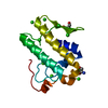
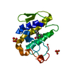
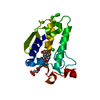
| Title | Crystal Structure of Porcine Pancreatic Phospholipase A2 in Complex with Cholate | ||||||
 Components Components | Phospholipase A2, major isoenzyme | ||||||
 Keywords Keywords | HYDROLASE / Bile salt / cholate / CARBOXYLIC ESTER HYDROLASE / PLA2 / Pancreatic Enzyme | ||||||
| Function / homology |  Function and homology information Function and homology informationregulation of D-glucose import across plasma membrane / positive regulation of podocyte apoptotic process / phosphatidylglycerol metabolic process / phosphatidylcholine metabolic process / leukotriene biosynthetic process / A2-type glycerophospholipase activity / : / bile acid binding / phospholipase A2 / neutrophil mediated immunity ...regulation of D-glucose import across plasma membrane / positive regulation of podocyte apoptotic process / phosphatidylglycerol metabolic process / phosphatidylcholine metabolic process / leukotriene biosynthetic process / A2-type glycerophospholipase activity / : / bile acid binding / phospholipase A2 / neutrophil mediated immunity / positive regulation of calcium ion transport into cytosol / lipid catabolic process / neutrophil chemotaxis / positive regulation of interleukin-8 production / phospholipid binding / positive regulation of immune response / positive regulation of fibroblast proliferation / fatty acid biosynthetic process / cellular response to insulin stimulus / positive regulation of MAPK cascade / intracellular signal transduction / signaling receptor binding / calcium ion binding / positive regulation of cell population proliferation / cell surface / positive regulation of transcription by RNA polymerase II / extracellular region Similarity search - Function | ||||||
| Biological species |  | ||||||
| Method |  X-RAY DIFFRACTION / MOLECULAR REPLACEMENT / Resolution: 1.9 Å X-RAY DIFFRACTION / MOLECULAR REPLACEMENT / Resolution: 1.9 Å | ||||||
 Authors Authors | Pan, Y.H. / Bahnson, B.J. / Jain, M.K. | ||||||
 Citation Citation | Journal: J.Mol.Biol. / Year: 2007 Title: Structural basis for bile salt inhibition of pancreatic phospholipase A2. Authors: Pan, Y.H. / Bahnson, B.J. | ||||||
| History |
|
- Structure visualization
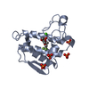
| Structure viewer | Molecule: MolmilJmol/JSmol |
|---|
- Downloads & links
-Download
| PDBx/mmCIF format | 2azy.cif.gz | 43.7 KB | Display | PDBx/mmCIF format |
|---|---|---|---|---|
| PDB format | pdb2azy.ent.gz | 29.2 KB | Display | PDB format |
| PDBx/mmJSON format | 2azy.json.gz | Tree view | PDBx/mmJSON format | |
| Others |  Other downloads Other downloads |
-Validation report
| Arichive directory | https://data.pdbj.org/pub/pdb/validation_reports/az/2azyftp://data.pdbj.org/pub/pdb/validation_reports/az/2azy | HTTPS FTP |
|---|
-Related structure data
| Related structure data |  2azzC 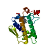 2b00C  2b01C  2b03C  2b04C  2bo1S C: citing same article ( S: Starting model for refinement |
|---|---|
| Similar structure data |


-Links
 PDBj
PDBj

- Assembly
| Deposited unit | 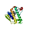
| ||||||||||||
|---|---|---|---|---|---|---|---|---|---|---|---|---|---|
| 1 |
| ||||||||||||
| 2 | 
| ||||||||||||
| Unit cell |
| ||||||||||||
| Components on special symmetry positions |
|
-Components
| #1: Protein | Mass: 14009.714 Da / Num. of mol.: 1 Source method: isolated from a genetically manipulated source Source: (gene. exp.)  |
|---|---|
| #2: Chemical | ChemComp-CA /   Mass: 40.078 Da / Num. of mol.: 1 / Source method: obtained synthetically / Formula: Ca Mass: 40.078 Da / Num. of mol.: 1 / Source method: obtained synthetically / Formula: Ca |
| #3: Chemical | ChemComp-CHD / 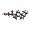  Mass: 408.571 Da / Num. of mol.: 1 / Source method: obtained synthetically / Formula: C24H40O5 Mass: 408.571 Da / Num. of mol.: 1 / Source method: obtained synthetically / Formula: C24H40O5 |
| #4: Water | ChemComp-HOH /  Mass: 18.015 Da / Num. of mol.: 193 / Source method: isolated from a natural source / Formula: H2O Mass: 18.015 Da / Num. of mol.: 193 / Source method: isolated from a natural source / Formula: H2O |
| Has protein modification | Y |
-Experimental details
-Experiment
| Experiment | Method: X-RAY DIFFRACTION / Number of used crystals: 1 |
|---|
- Sample preparation
| Crystal | Density Matthews: 3.26 Å3/Da / Density % sol: 62.22 % |
|---|---|
| Crystal grow | Temperature: 298 K / Method: vapor diffusion / pH: 8.5 / Details: 8%PEG8K, pH 8.5, VAPOR DIFFUSION, temperature 298K |
-Data collection
| Diffraction | Mean temperature: 93 K |
|---|---|
| Diffraction source | Source: ROTATING ANODE / Type: RIGAKU RU300 / Wavelength: 1.5418 Å |
| Detector | Type: RIGAKU RAXIS IV / Detector: IMAGE PLATE / Date: Dec 3, 2004 / Details: mirrors |
| Radiation | Monochromator: yale mirror / Protocol: SINGLE WAVELENGTH / Monochromatic (M) / Laue (L): M / Scattering type: x-ray |
| Radiation wavelength | Wavelength: 1.5418 Å / Relative weight: 1 |
| Reflection | Resolution: 1.9→35 Å / Num. obs: 14784 / % possible obs: 96.3 % / Observed criterion σ(F): 0 / Observed criterion σ(I): -3 / Redundancy: 4 % / Rmerge(I) obs: 0.049 / Rsym value: 0.038 / Χ2: 1.5 / Net I/σ(I): 33 |
| Reflection shell | Resolution: 1.9→1.97 Å / % possible obs: 93.6 % / Redundancy: 5 % / Rmerge(I) obs: 0.296 / Mean I/σ(I) obs: 6 / Num. measured obs: 1333 / Rsym value: 0.255 / Χ2: 1.25 / % possible all: 93.6 |
- Processing
| Software |
| |||||||||||||||||||||||||
|---|---|---|---|---|---|---|---|---|---|---|---|---|---|---|---|---|---|---|---|---|---|---|---|---|---|---|
| Refinement | Method to determine structure: MOLECULAR REPLACEMENT Starting model: PDB entry 2BO1 Resolution: 1.9→35 Å / Cross valid method: THROUGHT / σ(F): 0 / Stereochemistry target values: Engh & Huber
| |||||||||||||||||||||||||
| Refine analyze |
| |||||||||||||||||||||||||
| Refinement step | Cycle: LAST / Resolution: 1.9→35 Å
| |||||||||||||||||||||||||
| Refine LS restraints |
| |||||||||||||||||||||||||
| Xplor file |
|
