 Movie
Movie Controller


+ Open data

- Basic information
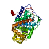
| Entry | Database: PDB / ID: 2ae1 | ||||||
|---|---|---|---|---|---|---|---|
| Title | TROPINONE REDUCTASE-II | ||||||
 Components Components | TROPINONE REDUCTASE-II | ||||||
 Keywords Keywords | OXIDOREDUCTASE / TROPANE ALKALOID BIOSYNTHESIS / REDUCTION OF TROPINONE TO PSEUDOTROPINE / SHORT-CHAIN DEHYDROGENASE | ||||||
| Function / homology |  Function and homology information Function and homology informationtropinone reductase II / tropinone reductase activity / tropane alkaloid biosynthetic process Similarity search - Function | ||||||
| Biological species |  Datura stramonium (jimsonweed) Datura stramonium (jimsonweed) | ||||||
| Method |  X-RAY DIFFRACTION / MIRAS / Resolution: 2.3 Å X-RAY DIFFRACTION / MIRAS / Resolution: 2.3 Å | ||||||
 Authors Authors | Nakajima, K. / Yamashita, A. / Akama, H. / Nakatsu, T. / Kato, H. / Hashimoto, T. / Oda, J. / Yamada, Y. | ||||||
 Citation Citation | Journal: Proc.Natl.Acad.Sci.USA / Year: 1998 Title: Crystal structures of two tropinone reductases: different reaction stereospecificities in the same protein fold. Authors: Nakajima, K. / Yamashita, A. / Akama, H. / Nakatsu, T. / Kato, H. / Hashimoto, T. / Oda, J. / Yamada, Y. #1: Journal: Proc.Natl.Acad.Sci.USA / Year: 1993Title: Two Tropinone Reductases with Different Stereospecificities are Short-Chain Dehydrogenases Evolved from a Common Ancestor Authors: Nakajima, K. / Hashimoto, T. / Yamada, Y. | ||||||
| History |
|
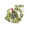
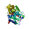
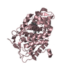
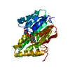
- Structure visualization
| Structure viewer | Molecule: MolmilJmol/JSmol |
|---|
- Downloads & links
-Download
| PDBx/mmCIF format | 2ae1.cif.gz | 57.8 KB | Display | PDBx/mmCIF format |
|---|---|---|---|---|
| PDB format | pdb2ae1.ent.gz | 46 KB | Display | PDB format |
| PDBx/mmJSON format | 2ae1.json.gz | Tree view | PDBx/mmJSON format | |
| Others |  Other downloads Other downloads |
-Validation report
| Summary document | 2ae1_validation.pdf.gz | 416 KB | Display | wwPDB validaton report |
|---|---|---|---|---|
| Full document | 2ae1_full_validation.pdf.gz | 419.3 KB | Display | |
| Data in XML | 2ae1_validation.xml.gz | 12.3 KB | Display | |
| Data in CIF | 2ae1_validation.cif.gz | 16.7 KB | Display | |
| Arichive directory | https://data.pdbj.org/pub/pdb/validation_reports/ae/2ae1ftp://data.pdbj.org/pub/pdb/validation_reports/ae/2ae1 | HTTPS FTP |
-Related structure data


-Links
 PDBj
PDBj


- Assembly
| Deposited unit | 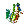
| ||||||||
|---|---|---|---|---|---|---|---|---|---|
| 1 |
| ||||||||
| 2 | 
| ||||||||
| Unit cell |
|
-Components
| #1: Protein | Mass: 28339.445 Da / Num. of mol.: 1 Source method: isolated from a genetically manipulated source Source: (gene. exp.) Datura stramonium (jimsonweed) / Cell line: BL21 / Organ: CULTURED ROOT / Plasmid: PETTR2 / Species (production host): Escherichia coli / Production host:  |
|---|---|
| #2: Water | ChemComp-HOH /  Mass: 18.015 Da / Num. of mol.: 103 / Source method: isolated from a natural source / Formula: H2O Mass: 18.015 Da / Num. of mol.: 103 / Source method: isolated from a natural source / Formula: H2O |
-Experimental details
-Experiment
| Experiment | Method: X-RAY DIFFRACTION / Number of used crystals: 1 |
|---|
- Sample preparation
| Crystal | Density Matthews: 2.24 Å3/Da / Density % sol: 45 % | ||||||||||||||||||||||||||||||||||||||||
|---|---|---|---|---|---|---|---|---|---|---|---|---|---|---|---|---|---|---|---|---|---|---|---|---|---|---|---|---|---|---|---|---|---|---|---|---|---|---|---|---|---|
| Crystal grow | pH: 6 / Details: pH 6.0 | ||||||||||||||||||||||||||||||||||||||||
| Crystal | *PLUS | ||||||||||||||||||||||||||||||||||||||||
| Crystal grow | *PLUS Temperature: 20 ℃ / pH: 7.5 / Method: vapor diffusion, hanging drop | ||||||||||||||||||||||||||||||||||||||||
| Components of the solutions | *PLUS
|
-Data collection
| Diffraction | Mean temperature: 113 K |
|---|---|
| Diffraction source | Source: ROTATING ANODE / Type: RIGAKU RUH3R / Wavelength: 1.5418 |
| Detector | Type: RIGAKU / Detector: IMAGE PLATE / Date: Sep 7, 1996 / Details: COLLIMATOR |
| Radiation | Monochromator: GRAPHITE(002) / Monochromatic (M) / Laue (L): M / Scattering type: x-ray |
| Radiation wavelength | Wavelength: 1.5418 Å / Relative weight: 1 |
| Reflection | Resolution: 2.03→64.2 Å / Num. obs: 14339 / % possible obs: 81.8 % / Observed criterion σ(I): 1 / Redundancy: 7.2 % / Rmerge(I) obs: 0.087 / Net I/σ(I): 11.65 |
| Reflection shell | Resolution: 2.02→2.25 Å / Rmerge(I) obs: 0.216 / Mean I/σ(I) obs: 2.81 / % possible all: 55 |
| Reflection | *PLUS Num. measured all: 103356 |
- Processing
| Software |
| ||||||||||||||||||||||||||||||||||||||||||||||||||||||||||||
|---|---|---|---|---|---|---|---|---|---|---|---|---|---|---|---|---|---|---|---|---|---|---|---|---|---|---|---|---|---|---|---|---|---|---|---|---|---|---|---|---|---|---|---|---|---|---|---|---|---|---|---|---|---|---|---|---|---|---|---|---|---|
| Refinement | Method to determine structure: MIRAS / Resolution: 2.3→10 Å / Cross valid method: THROUGHOUT / σ(F): 2
| ||||||||||||||||||||||||||||||||||||||||||||||||||||||||||||
| Refine analyze |
| ||||||||||||||||||||||||||||||||||||||||||||||||||||||||||||
| Refinement step | Cycle: LAST / Resolution: 2.3→10 Å
| ||||||||||||||||||||||||||||||||||||||||||||||||||||||||||||
| Refine LS restraints |
| ||||||||||||||||||||||||||||||||||||||||||||||||||||||||||||
| LS refinement shell | Resolution: 2.3→2.4 Å / Total num. of bins used: 8
| ||||||||||||||||||||||||||||||||||||||||||||||||||||||||||||
| Xplor file |
| ||||||||||||||||||||||||||||||||||||||||||||||||||||||||||||
| Software | *PLUS Name: X-PLOR / Version: 3.851 / Classification: refinement | ||||||||||||||||||||||||||||||||||||||||||||||||||||||||||||
| Refine LS restraints | *PLUS
|
