 Movie
Movie Controller

[English] 日本語
 Yorodumi
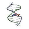
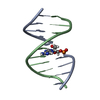
- PDB-208d: HIGH-RESOLUTION STRUCTURE OF A DNA HELIX FORMING (C.G)*G BASE TRIPLETS -
+ Open data

- Basic information
| Entry | Database: PDB / ID: 208d | ||||||||||||||||||
|---|---|---|---|---|---|---|---|---|---|---|---|---|---|---|---|---|---|---|---|
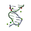
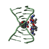
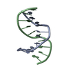
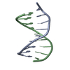
| Title | HIGH-RESOLUTION STRUCTURE OF A DNA HELIX FORMING (C.G)*G BASE TRIPLETS | ||||||||||||||||||
 Components Components | DNA (5'-D(* Keywords KeywordsDNA / B-DNA / DOUBLE HELIX / BASE TRIPLET | Function / homology | DNA |  Function and homology information Function and homology informationMethod |  X-RAY DIFFRACTION / MOLECULAR REPLACEMENT / Resolution: 2.05 Å X-RAY DIFFRACTION / MOLECULAR REPLACEMENT / Resolution: 2.05 Å  Authors AuthorsVan Meervelt, L. / Vlieghe, D. / Dautant, A. / Gallois, B. / Precigoux, G. / Kennard, O. |  CitationJournal: Nature / Year: 1995 CitationJournal: Nature / Year: 1995Title: High-resolution structure of a DNA helix forming (C.G)*G base triplets. Authors: Van Meervelt, L. / Vlieghe, D. / Dautant, A. / Gallois, B. / Precigoux, G. / Kennard, O. #1: Journal: Acta Crystallogr.,Sect.D / Year: 1996Title: Formation of (C.G)*G Triplets in a B-DNA Duplex with Overhanging Bases Authors: Vlieghe, D. / Van Meervelt, L. / Dautant, A. / Gallois, B. / Precigoux, G. / Kennard, O. History |
|
- Structure visualization
| Structure viewer | Molecule: MolmilJmol/JSmol |
|---|
- Downloads & links
-Download
| PDBx/mmCIF format | 208d.cif.gz | 21.2 KB | Display | PDBx/mmCIF format |
|---|---|---|---|---|
| PDB format | pdb208d.ent.gz | 13.3 KB | Display | PDB format |
| PDBx/mmJSON format | 208d.json.gz | Tree view | PDBx/mmJSON format | |
| Others |  Other downloads Other downloads |
-Validation report
| Arichive directory | https://data.pdbj.org/pub/pdb/validation_reports/08/208dftp://data.pdbj.org/pub/pdb/validation_reports/08/208d | HTTPS FTP |
|---|
-Related structure data
| Similar structure data |
|---|


-Links
 PDBj
PDBj

- Assembly
| Deposited unit | 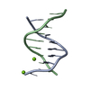
| ||||||||
|---|---|---|---|---|---|---|---|---|---|
| 1 |
| ||||||||
| Unit cell |
|
-Components
| #1: DNA chain | Mass: 2755.823 Da / Num. of mol.: 2 / Source method: obtained synthetically #2: Chemical |   Mass: 24.305 Da / Num. of mol.: 2 / Source method: obtained synthetically / Formula: Mg Mass: 24.305 Da / Num. of mol.: 2 / Source method: obtained synthetically / Formula: Mg#3: Water | ChemComp-HOH / |  Mass: 18.015 Da / Num. of mol.: 86 / Source method: isolated from a natural source / Formula: H2O Mass: 18.015 Da / Num. of mol.: 86 / Source method: isolated from a natural source / Formula: H2O |
|---|
-Experimental details
-Experiment
| Experiment | Method: X-RAY DIFFRACTION |
|---|
- Sample preparation
| Crystal | Density Matthews: 1.94 Å3/Da / Density % sol: 46 % | ||||||||||||||||||||||||||||||||||||||||||
|---|---|---|---|---|---|---|---|---|---|---|---|---|---|---|---|---|---|---|---|---|---|---|---|---|---|---|---|---|---|---|---|---|---|---|---|---|---|---|---|---|---|---|---|
| Crystal grow | Temperature: 289 K / Method: vapor diffusion / pH: 6 / Details: pH 6.00, VAPOR DIFFUSION, temperature 289.00K | ||||||||||||||||||||||||||||||||||||||||||
| Components of the solutions |
| ||||||||||||||||||||||||||||||||||||||||||
| Crystal | *PLUS | ||||||||||||||||||||||||||||||||||||||||||
| Crystal grow | *PLUS Temperature: 16 ℃ / pH: 6 | ||||||||||||||||||||||||||||||||||||||||||
| Components of the solutions | *PLUS
|
-Data collection
| Diffraction | Mean temperature: 291 K |
|---|---|
| Diffraction source | Source: ROTATING ANODE |
| Detector | Type: MARRESEARCH / Detector: IMAGE PLATE / Date: May 2, 1994 |
| Radiation | Monochromatic (M) / Laue (L): M / Scattering type: x-ray |
| Radiation wavelength | Relative weight: 1 |
| Reflection | Num. all: 22402 / Num. obs: 2824 / % possible obs: 87 % / Observed criterion σ(I): 3 / Redundancy: 7.9 % / Rmerge(I) obs: 0.036 |
| Reflection | *PLUS Highest resolution: 2.05 Å / % possible obs: 87 % / Observed criterion σ(I): 3 |
- Processing
| Software |
| ||||||||||||||||||||||||||||||||||||||||||||||||||||||||||||
|---|---|---|---|---|---|---|---|---|---|---|---|---|---|---|---|---|---|---|---|---|---|---|---|---|---|---|---|---|---|---|---|---|---|---|---|---|---|---|---|---|---|---|---|---|---|---|---|---|---|---|---|---|---|---|---|---|---|---|---|---|---|
| Refinement | Method to determine structure: MOLECULAR REPLACEMENT Starting model: BDL001 Resolution: 2.05→8 Å / σ(F): 4
| ||||||||||||||||||||||||||||||||||||||||||||||||||||||||||||
| Refine Biso |
| ||||||||||||||||||||||||||||||||||||||||||||||||||||||||||||
| Refinement step | Cycle: LAST / Resolution: 2.05→8 Å
| ||||||||||||||||||||||||||||||||||||||||||||||||||||||||||||
| Refine LS restraints |
| ||||||||||||||||||||||||||||||||||||||||||||||||||||||||||||
| Software | *PLUS Name: X-PLOR / Classification: refinement | ||||||||||||||||||||||||||||||||||||||||||||||||||||||||||||
| Refinement | *PLUS Highest resolution: 2.05 Å / Lowest resolution: 8 Å / σ(F): 4 | ||||||||||||||||||||||||||||||||||||||||||||||||||||||||||||
| Solvent computation | *PLUS | ||||||||||||||||||||||||||||||||||||||||||||||||||||||||||||
| Displacement parameters | *PLUS Biso mean: 18.4 Å2 | ||||||||||||||||||||||||||||||||||||||||||||||||||||||||||||
| Refine LS restraints | *PLUS
|
