 Movie
Movie Controller


+ Open data

- Basic information
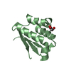
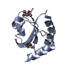
| Entry | Database: PDB / ID: 1zzy | ||||||
|---|---|---|---|---|---|---|---|
| Title | Crystal Structure of Thioredoxin Mutant L7V | ||||||
 Components Components | Thioredoxin 1 | ||||||
 Keywords Keywords | ELECTRON TRANSPORT / Alpha/Beta | ||||||
| Function / homology |  Function and homology information Function and homology informationpositive regulation of DNA-directed DNA polymerase activity / DNA polymerase processivity factor activity / protein-disulfide reductase activity / cell redox homeostasis / oxidoreductase activity / cytoplasm / cytosol Similarity search - Function | ||||||
| Biological species |  | ||||||
| Method |  X-RAY DIFFRACTION / MOLECULAR REPLACEMENT / Resolution: 2.5 Å X-RAY DIFFRACTION / MOLECULAR REPLACEMENT / Resolution: 2.5 Å | ||||||
 Authors Authors | Gavira, J.A. / Perez-Jimenez, R. / Ibarra-Molero, B. / Sanchez-Ruiz, J.M. | ||||||
 Citation Citation | Journal: To be Published Title: Crystal Structure of Thioredoxin Mutant L7V Authors: Gavira, J.A. / Perez-Jimenez, R. / Ibarra-Molero, B. / Sanchez-Ruiz, J.M. | ||||||
| History |
|
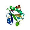
- Structure visualization
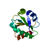
| Structure viewer | Molecule: MolmilJmol/JSmol |
|---|
- Downloads & links
-Download
| PDBx/mmCIF format | 1zzy.cif.gz | 51.6 KB | Display | PDBx/mmCIF format |
|---|---|---|---|---|
| PDB format | pdb1zzy.ent.gz | 36.7 KB | Display | PDB format |
| PDBx/mmJSON format | 1zzy.json.gz | Tree view | PDBx/mmJSON format | |
| Others |  Other downloads Other downloads |
-Validation report
| Summary document | 1zzy_validation.pdf.gz | 428.9 KB | Display | wwPDB validaton report |
|---|---|---|---|---|
| Full document | 1zzy_full_validation.pdf.gz | 430.8 KB | Display | |
| Data in XML | 1zzy_validation.xml.gz | 9.5 KB | Display | |
| Data in CIF | 1zzy_validation.cif.gz | 12.5 KB | Display | |
| Arichive directory | https://data.pdbj.org/pub/pdb/validation_reports/zz/1zzyftp://data.pdbj.org/pub/pdb/validation_reports/zz/1zzy | HTTPS FTP |
-Related structure data
| Related structure data | 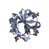 2trxS S: Starting model for refinement |
|---|---|
| Similar structure data |


-Links
 PDBj
PDBj

- Assembly
| Deposited unit | 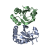
| ||||||||
|---|---|---|---|---|---|---|---|---|---|
| 1 | 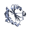
| ||||||||
| 2 | 
| ||||||||
| Unit cell |
|
-Components
| #1: Protein | Mass: 11673.360 Da / Num. of mol.: 2 / Mutation: L7V Source method: isolated from a genetically manipulated source Source: (gene. exp.) #2: Water | ChemComp-HOH / |  Mass: 18.015 Da / Num. of mol.: 35 / Source method: isolated from a natural source / Formula: H2O Mass: 18.015 Da / Num. of mol.: 35 / Source method: isolated from a natural source / Formula: H2O |
|---|
-Experimental details
-Experiment
| Experiment | Method: X-RAY DIFFRACTION / Number of used crystals: 1 |
|---|
- Sample preparation
| Crystal | Density Matthews: 2.08 Å3/Da / Density % sol: 40.99 % |
|---|---|
| Crystal grow | Temperature: 295 K / Method: counter-diffusion / pH: 3.8 Details: 10 mM AcNa pH 3.8, 25% (v/v) EtOH, 10 mM Ac2Cu, Counter-diffusion, temperature 295K |
-Data collection
| Diffraction | Mean temperature: 293 K |
|---|---|
| Diffraction source | Source: ROTATING ANODE / Type: BRUKER AXS MICROSTAR / Wavelength: 1.5418 Å |
| Detector | Type: BRUKER SMART 6000 / Detector: CCD / Date: Jan 7, 2005 / Details: Montel Optics |
| Radiation | Monochromator: Ni Filter / Protocol: SINGLE WAVELENGTH / Monochromatic (M) / Laue (L): M / Scattering type: x-ray |
| Radiation wavelength | Wavelength: 1.5418 Å / Relative weight: 1 |
| Reflection | Resolution: 2.5→40.26 Å / Num. all: 5671 / Num. obs: 5671 / % possible obs: 88.7 % / Redundancy: 1.46 % / Biso Wilson estimate: 12.8 Å2 / Limit h max: 13 / Limit h min: -12 / Limit k max: 14 / Limit k min: -12 / Limit l max: 17 / Limit l min: 0 / Observed criterion F max: 1312437.97 / Observed criterion F min: 3.46 / Rsym value: 0.1236 / Net I/σ(I): 6.57 |
| Reflection shell | Resolution: 2.5→2.6 Å / Redundancy: 1.38 % / Mean I/σ(I) obs: 2.82 / Num. unique all: 605 / Rsym value: 0.374 / % possible all: 85.2 |
- Processing
| Software |
| ||||||||||||||||||||||||||||||||||||||||||||||||||||||||||||||||||||||||||||||||||||||||||
|---|---|---|---|---|---|---|---|---|---|---|---|---|---|---|---|---|---|---|---|---|---|---|---|---|---|---|---|---|---|---|---|---|---|---|---|---|---|---|---|---|---|---|---|---|---|---|---|---|---|---|---|---|---|---|---|---|---|---|---|---|---|---|---|---|---|---|---|---|---|---|---|---|---|---|---|---|---|---|---|---|---|---|---|---|---|---|---|---|---|---|---|
| Refinement | Method to determine structure: MOLECULAR REPLACEMENT Starting model: A:2trx.pdb Resolution: 2.5→38.69 Å / Rfactor Rfree error: 0.016 / Occupancy max: 1 / Occupancy min: 0.1 / Isotropic thermal model: RESTRAINED / Cross valid method: THROUGHOUT / σ(F): 0 / Stereochemistry target values: Engh & Huber Details: XTALVIEW and MOLBROBITY were also used for the refinement.
| ||||||||||||||||||||||||||||||||||||||||||||||||||||||||||||||||||||||||||||||||||||||||||
| Solvent computation | Solvent model: CNS bulk solvent model used / Bsol: 50.8937 Å2 / ksol: 0.324627 e/Å3 | ||||||||||||||||||||||||||||||||||||||||||||||||||||||||||||||||||||||||||||||||||||||||||
| Displacement parameters | Biso max: 72.55 Å2 / Biso mean: 23.29 Å2 / Biso min: 1.01 Å2
| ||||||||||||||||||||||||||||||||||||||||||||||||||||||||||||||||||||||||||||||||||||||||||
| Refine analyze |
| ||||||||||||||||||||||||||||||||||||||||||||||||||||||||||||||||||||||||||||||||||||||||||
| Refinement step | Cycle: LAST / Resolution: 2.5→38.69 Å
| ||||||||||||||||||||||||||||||||||||||||||||||||||||||||||||||||||||||||||||||||||||||||||
| Refine LS restraints |
| ||||||||||||||||||||||||||||||||||||||||||||||||||||||||||||||||||||||||||||||||||||||||||
| LS refinement shell | Refine-ID: X-RAY DIFFRACTION / Total num. of bins used: 8
| ||||||||||||||||||||||||||||||||||||||||||||||||||||||||||||||||||||||||||||||||||||||||||
| Xplor file |
|
