 Movie
Movie Controller

[English] 日本語
 Yorodumi
- PDB-1ypt: CRYSTAL STRUCTURE OF YERSINIA PROTEIN TYROSINE PHOSPHATASE AT 2.5... -
+ Open data

- Basic information
| Entry | Database: PDB / ID: 1ypt | ||||||
|---|---|---|---|---|---|---|---|
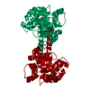
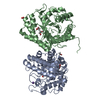
| Title | CRYSTAL STRUCTURE OF YERSINIA PROTEIN TYROSINE PHOSPHATASE AT 2.5 ANGSTROMS AND THE COMPLEX WITH TUNGSTATE | ||||||
 Components Components | PROTEIN-TYROSINE PHOSPHATASE YERSINIA (CATALYTIC DOMAIN) | ||||||
 Keywords Keywords |  HYDROLASE / PROTEIN TYROSINE PHOSPHATASE HYDROLASE / PROTEIN TYROSINE PHOSPHATASE | ||||||
| Function / homology |  Function and homology informationdephosphorylation / protein-tyrosine-phosphatase / protein tyrosine phosphatase activity / extracellular region Function and homology informationdephosphorylation / protein-tyrosine-phosphatase / protein tyrosine phosphatase activity / extracellular regionSimilarity search - Function | ||||||
| Biological species |  Yersinia enterocolitica (bacteria) Yersinia enterocolitica (bacteria) | ||||||
| Method | X-RAY DIFFRACTION / Resolution: 2.5 Å | ||||||
 Authors Authors | Stuckey, J.A. / Schubert, H.L. / Fauman, E.B. / Zhang, Z.-Y. / Dixon, J.E. / Saper, M.A. | ||||||
 Citation Citation | Journal: Nature / Year: 1994 Title: Crystal structure of Yersinia protein tyrosine phosphatase at 2.5 A and the complex with tungstate. Authors: Stuckey, J.A. / Schubert, H.L. / Fauman, E.B. / Zhang, Z.Y. / Dixon, J.E. / Saper, M.A. #1: Journal: J.Biol.Chem. / Year: 1992Title: Expression, Purification, and Physicochemical Characterization of a Recombinant Yersinia Protein Tyrosine Phosphatase Authors: Zhang, Z.-Y. / Clemens, J.C. / Schubert, H.L. / Stuckey, J.A. / Fischer, M.W.F. / Hume, D.M. / Saper, M.A. / Dixon, J.E. | ||||||
| History |
|
- Structure visualization
| Structure viewer | Molecule: MolmilJmol/JSmol |
|---|
- Downloads & links
-Download
| PDBx/mmCIF format | 1ypt.cif.gz | 118.4 KB | Display | PDBx/mmCIF format |
|---|---|---|---|---|
| PDB format | pdb1ypt.ent.gz | 91.9 KB | Display | PDB format |
| PDBx/mmJSON format | 1ypt.json.gz | Tree view | PDBx/mmJSON format | |
| Others |  Other downloads Other downloads |
-Validation report
| Arichive directory | https://data.pdbj.org/pub/pdb/validation_reports/yp/1yptftp://data.pdbj.org/pub/pdb/validation_reports/yp/1ypt | HTTPS FTP |
|---|
-Related structure data
| Similar structure data |
|---|


-Links
 PDBj
PDBj


- Assembly
| Deposited unit | 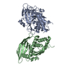
| ||||||||
|---|---|---|---|---|---|---|---|---|---|
| 1 |
| ||||||||
| Unit cell |
| ||||||||
| Noncrystallographic symmetry (NCS) | NCS oper: (Code: given Matrix: (0.9922, 0.091653, 0.08449), Vector : Details | MTRIX THE TRANSFORMATIONS PRESENTED ON MTRIX RECORDS BELOW DESCRIBE NON-CRYSTALLOGRAPHIC RELATIONSHIPS AMONG THE VARIOUS DOMAINS IN THIS ENTRY. APPLYING THE APPROPRIATE MTRIX TRANSFORMATION TO THE RESIDUES LISTED FIRST WILL YIELD APPROXIMATE COORDINATES FOR THE RESIDUES LISTED SECOND. APPLIED TO TRANSFORMED TO MTRIX CHAIN RESIDUES CHAIN RESIDUES RMSD M1 A 191 - A 468 B 191 - B 468 .223 A 178.02 DEGREE ROTATION ABOUT THE X AXIS OF THE CRYSTAL THAT RELATES MOLECULE A TO MOLECULE B. THE RMSD (EXCLUDING 4 FLEXIBLE LOOPS) BETWEEN MOLECULE A AND MOLECULE B IS 0.196. THE RMSD USING ALL ATOMS IS 0.523. | |
-Components
| #1: Protein | Mass: 33422.688 Da / Num. of mol.: 2 Source method: isolated from a genetically manipulated source Source: (gene. exp.) Yersinia enterocolitica (bacteria) / Gene: YOP51 / Plasmid: PT7-7 GENE: YOP51 / Gene (production host): YOP51 / References: UniProt: P15273, protein-tyrosine-phosphatase#2: Water | ChemComp-HOH / | Water Mass: 18.015 Da / Num. of mol.: 65 / Source method: isolated from a natural source / Formula: H2O Mass: 18.015 Da / Num. of mol.: 65 / Source method: isolated from a natural source / Formula: H2O |
|---|
-Experimental details
-Experiment
| Experiment | Method: X-RAY DIFFRACTION |
|---|
- Sample preparation
| Crystal | Density Matthews: 2.37 Å3/Da / Density % sol: 48.19 % | ||||||||||||||||||||||||||||||||||||||||||
|---|---|---|---|---|---|---|---|---|---|---|---|---|---|---|---|---|---|---|---|---|---|---|---|---|---|---|---|---|---|---|---|---|---|---|---|---|---|---|---|---|---|---|---|
| Crystal grow | Details: THE CATALYTIC DOMAIN (RESIDUES 163 - 468) OF YOP51 WAS CRYSTALLIZED AT 22 DEGREES CELSIUS, IN A SOLUTION OF 10 - 16% POLYETHYLENE GLYCOL, 5% 2-METHYL-2,4-PENTANEDIOL, 0.1% B-MERCAPTOETHANOL, ...Details: THE CATALYTIC DOMAIN (RESIDUES 163 - 468) OF YOP51 WAS CRYSTALLIZED AT 22 DEGREES CELSIUS, IN A SOLUTION OF 10 - 16% POLYETHYLENE GLYCOL, 5% 2-METHYL-2,4-PENTANEDIOL, 0.1% B-MERCAPTOETHANOL, AND 1MM IMIDAZOLE, PH 7.2. SOURCE ONLY THE CATALYTIC DOMAIN (RESIDUES 163 - 468) WAS EXPRESSED FOR THIS PARTICULAR EXPERIMENT. THE 235 CYS TO ARG MUTATION WAS UNINTENTIONAL AND MOST LIKELY THE RESULT OF USING PCR TO CONSTRUCT THE PLASMID (SEE REFERENCE REMARK 1). | ||||||||||||||||||||||||||||||||||||||||||
| Crystal | *PLUS Density % sol: 49 % | ||||||||||||||||||||||||||||||||||||||||||
| Crystal grow | *PLUS Temperature: 22 ℃ / pH: 7.2 / Method: vapor diffusion, hanging drop | ||||||||||||||||||||||||||||||||||||||||||
| Components of the solutions | *PLUS
|
-Data collection
| Reflection | Num. obs: 19476 / % possible obs: 91 % / Observed criterion σ(I): 0 |
|---|---|
| Reflection | *PLUS Highest resolution: 2.5 Å / Rmerge(I) obs: 0.059 |
- Processing
| Software |
| ||||||||||||||||||||||||||||||||||||||||||||||||||||||||||||
|---|---|---|---|---|---|---|---|---|---|---|---|---|---|---|---|---|---|---|---|---|---|---|---|---|---|---|---|---|---|---|---|---|---|---|---|---|---|---|---|---|---|---|---|---|---|---|---|---|---|---|---|---|---|---|---|---|---|---|---|---|---|
| Refinement | Resolution: 2.5→7 Å / σ(F): 2
| ||||||||||||||||||||||||||||||||||||||||||||||||||||||||||||
| Refinement step | Cycle: LAST / Resolution: 2.5→7 Å
| ||||||||||||||||||||||||||||||||||||||||||||||||||||||||||||
| Refine LS restraints |
| ||||||||||||||||||||||||||||||||||||||||||||||||||||||||||||
| Software | *PLUS Name: X-PLOR / Classification: refinement | ||||||||||||||||||||||||||||||||||||||||||||||||||||||||||||
| Refinement | *PLUS Rfactor obs: 0.168 / Rfactor Rfree: 0.222 | ||||||||||||||||||||||||||||||||||||||||||||||||||||||||||||
| Solvent computation | *PLUS | ||||||||||||||||||||||||||||||||||||||||||||||||||||||||||||
| Displacement parameters | *PLUS | ||||||||||||||||||||||||||||||||||||||||||||||||||||||||||||
| Refine LS restraints | *PLUS
|
