 Movie
Movie Controller

[English] 日本語
 Yorodumi
- PDB-3qz6: The crystal structure of HpcH/HpaI aldolase from Desulfitobacteri... -
+ Open data

- Basic information
| Entry | Database: PDB / ID: 3qz6 | ||||||
|---|---|---|---|---|---|---|---|
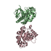
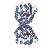
| Title | The crystal structure of HpcH/HpaI aldolase from Desulfitobacterium hafniense DCB-2 | ||||||
 Components Components | HpcH/HpaI aldolase | ||||||
 Keywords Keywords | LYASE / structural genomics / PSI-Biology / protein structure initiative / midwest center for structural genomics / MCSG / cytoplasmic | ||||||
| Function / homology |  Function and homology information Function and homology information | ||||||
| Biological species |  Desulfitobacterium hafniense (bacteria) Desulfitobacterium hafniense (bacteria) | ||||||
| Method |  X-RAY DIFFRACTION / SYNCHROTRON / SAD / Resolution: 1.999 Å X-RAY DIFFRACTION / SYNCHROTRON / SAD / Resolution: 1.999 Å | ||||||
 Authors Authors | Tan, K. / Chhor, G. / Clancy, S. / Joachimiak, A. / Midwest Center for Structural Genomics (MCSG) | ||||||
 Citation Citation | Journal: To be Published Title: The crystal structure of HpcH/HpaI aldolase from Desulfitobacterium hafniense DCB-2 Authors: Tan, K. / Chhor, G. / Clancy, S. / Joachimiak, A. | ||||||
| History |
|
- Structure visualization
| Structure viewer | Molecule: MolmilJmol/JSmol |
|---|
- Downloads & links
-Download
| PDBx/mmCIF format | 3qz6.cif.gz | 117.3 KB | Display | PDBx/mmCIF format |
|---|---|---|---|---|
| PDB format | pdb3qz6.ent.gz | 90.8 KB | Display | PDB format |
| PDBx/mmJSON format | 3qz6.json.gz | Tree view | PDBx/mmJSON format | |
| Others |  Other downloads Other downloads |
-Validation report
| Arichive directory | https://data.pdbj.org/pub/pdb/validation_reports/qz/3qz6ftp://data.pdbj.org/pub/pdb/validation_reports/qz/3qz6 | HTTPS FTP |
|---|
-Related structure data
| Similar structure data | |
|---|---|
| Other databases |


-Links
 PDBj
PDBj
- Assembly
| Deposited unit | 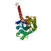
| ||||||||
|---|---|---|---|---|---|---|---|---|---|
| 1 | 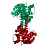
| ||||||||
| 2 | 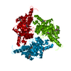
| ||||||||
| 3 | x 6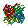
| ||||||||
| Unit cell |
| ||||||||
| Components on special symmetry positions |
|
-Components
| #1: Protein | Mass: 29090.256 Da / Num. of mol.: 1 Source method: isolated from a genetically manipulated source Source: (gene. exp.) Desulfitobacterium hafniense (bacteria)Strain: DCB-2 / Gene: Dhaf_2073 / Plasmid: pMCSG7 / Production host: |
|---|---|
| #2: Chemical | ChemComp-ZN /   Mass: 65.409 Da / Num. of mol.: 1 / Source method: obtained synthetically / Formula: Zn Mass: 65.409 Da / Num. of mol.: 1 / Source method: obtained synthetically / Formula: Zn |
| #3: Water | ChemComp-HOH /  Mass: 18.015 Da / Num. of mol.: 242 / Source method: isolated from a natural source / Formula: H2O Mass: 18.015 Da / Num. of mol.: 242 / Source method: isolated from a natural source / Formula: H2O |
| Has protein modification | Y |
-Experimental details
-Experiment
| Experiment | Method: X-RAY DIFFRACTION / Number of used crystals: 1 |
|---|
- Sample preparation
| Crystal grow | Temperature: 277 K / Method: vapor diffusion, sitting drop / pH: 8.5 Details: 0.2M Sodium citrate, 0.1M Tris.HCl, 15% (v/v) PEG400, pH 8.5, VAPOR DIFFUSION, SITTING DROP, temperature 277K |
|---|
-Data collection
| Diffraction | Mean temperature: 100 K |
|---|---|
| Diffraction source | Source: SYNCHROTRON / Site: APS  / Beamline: 19-ID / Wavelength: 0.97915 Å / Beamline: 19-ID / Wavelength: 0.97915 Å |
| Detector | Type: ADSC QUANTUM 315r / Detector: CCD / Date: Dec 6, 2010 / Details: Mirror |
| Radiation | Monochromator: Si 111 crystal / Protocol: SINGLE WAVELENGTH / Monochromatic (M) / Laue (L): M / Scattering type: x-ray |
| Radiation wavelength | Wavelength: 0.97915 Å / Relative weight: 1 |
| Reflection | Resolution: 2→44 Å / Num. all: 39752 / Num. obs: 39752 / % possible obs: 100 % / Observed criterion σ(F): 0 / Observed criterion σ(I): 0 / Redundancy: 5.5 % / Rmerge(I) obs: 0.773 / Net I/σ(I): 16 |
| Reflection shell | Resolution: 2→2.03 Å / Redundancy: 5.5 % / Rmerge(I) obs: 0.773 / Mean I/σ(I) obs: 0.773 / Num. unique all: 1931 / Rsym value: 0.016 / % possible all: 99.9 |
- Processing
| Software |
| |||||||||||||||||||||||||||||||||||||||||||||||||||||||||||||||||||||||||||||
|---|---|---|---|---|---|---|---|---|---|---|---|---|---|---|---|---|---|---|---|---|---|---|---|---|---|---|---|---|---|---|---|---|---|---|---|---|---|---|---|---|---|---|---|---|---|---|---|---|---|---|---|---|---|---|---|---|---|---|---|---|---|---|---|---|---|---|---|---|---|---|---|---|---|---|---|---|---|---|
| Refinement | Method to determine structure: SAD / Resolution: 1.999→43.828 Å / SU ML: 0.22 / σ(F): 0 / Phase error: 16.51 / Stereochemistry target values: ML
| |||||||||||||||||||||||||||||||||||||||||||||||||||||||||||||||||||||||||||||
| Solvent computation | Shrinkage radii: 0.95 Å / VDW probe radii: 1.2 Å / Solvent model: FLAT BULK SOLVENT MODEL / Bsol: 38.947 Å2 / ksol: 0.344 e/Å3 | |||||||||||||||||||||||||||||||||||||||||||||||||||||||||||||||||||||||||||||
| Displacement parameters |
| |||||||||||||||||||||||||||||||||||||||||||||||||||||||||||||||||||||||||||||
| Refinement step | Cycle: LAST / Resolution: 1.999→43.828 Å
| |||||||||||||||||||||||||||||||||||||||||||||||||||||||||||||||||||||||||||||
| Refine LS restraints |
| |||||||||||||||||||||||||||||||||||||||||||||||||||||||||||||||||||||||||||||
| LS refinement shell |
| |||||||||||||||||||||||||||||||||||||||||||||||||||||||||||||||||||||||||||||
| Refinement TLS params. | Method: refined / Origin x: 16.2071 Å / Origin y: 11.587 Å / Origin z: 17.1328 Å
| |||||||||||||||||||||||||||||||||||||||||||||||||||||||||||||||||||||||||||||
| Refinement TLS group | Selection details: chain A |
